Figures & data
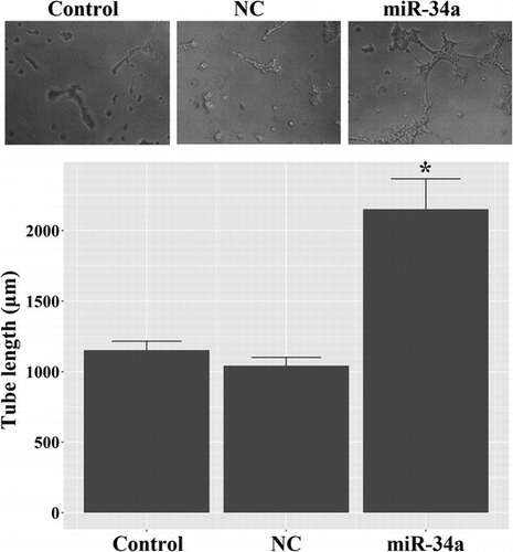
Fig. 1. Identification of the stem cell features of CD133 + U251 cells by immunofluorescent assay. CD133 + cells were isolated from U251 cells using anti-CD133 immunomagnetic beads, and the sorted cells were positively stained for CD133 and Nestin, representing stem cell-like characteristics. (A) representative images of immunofluorescent staining of CD133 and Nestin in the sorted cells. (B) quantitative analysis of the proportions of CD133 + and Nestin + cells in the sorted population. Scale bar, 50 μm.
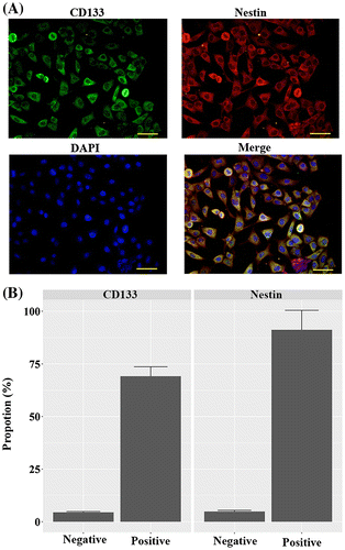
Fig. 2. Tube formation assay in CD133 + U251 cells transfected with miR-34a mimic or NC oligos. Up-regulation of miR-34a strikingly induced tube formation in CD133 + U251 cells. Magnification, 100 × . “*”, significantly different from Control and NC cells, p < 0.05.
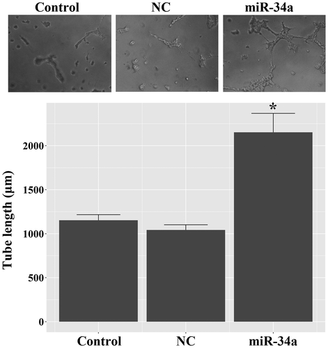
Fig. 3. Transfection with miR-34a mimics induced the expression of endothelial markers in CD133 + U251 cells. (A) Representative images of RT-PCR validation of the expression of endothelial markers: the expressions of CD31, CD34, and vWF were observed only in the CD133 + U251 cells transfected with miR-34a. (B) Representative images of western blotting validation of expressions of endothelial markers: the expressions of CD31, CD34, and vWF were observed only in the CD133 + U251 cells transfected with miR-34a. (C) Representative images of immunofluorescent staining of the three endothelial markers: more immunofluorescent positive cells were observed in CD133 + U251 cells transfected with miR-34a. Scale bar, 50 μm.
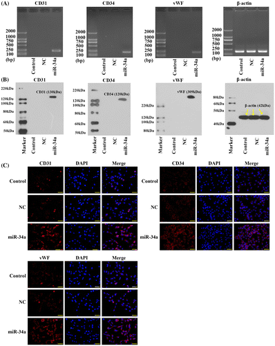
Fig. 4. Transfection with miR-34a mimics down-regulated Notch pathway components in CD133 + U251 cells. (A) Quantitative RT2-PCR analysis of the expression levels of Notch 1 and Dll1. (B) Representative images and quantitative analysis of western blotting assay of the expressions of Notch 1 and Dll1. “*” represents a significant difference from the Control and NC groups, p < 0.05.
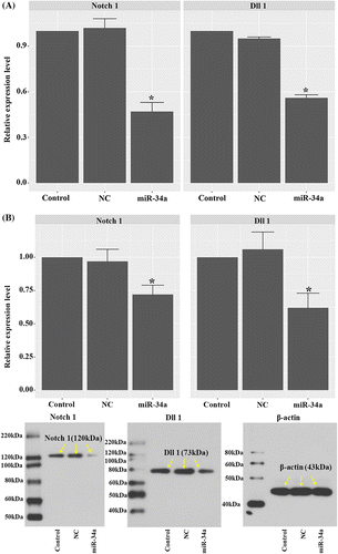
Fig. 5. Transfection with miR-34a mimics inhibited the expressions of Notch 1 and Dll1 as validated by immunofluorescent assay. (A) Representative images of immunofluorescent staining of Notch 1 in CD133 + U251 cells transfected with miR-34a mimics or NC oligos. (B) Representative images of immunofluorescent staining of Dll1 in different groups. Scale bar, 50 μm.
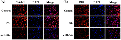
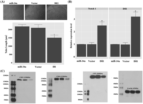
Fig. 6. Overexpression of Dll1 confounded the effect of miR-34a mimics to induce vasculature transdifferentiation of CD133 + U251 cells by inducing the expression of Notch 1. (A) Overexpression of Dll1 significantly inhibited tube formation in miR-34a mimics transfected CD133 + U251 cells. 100×. (B) Quantitative RT2-PCR analysis of the expression levels of Notch 1 and Dll1. (C) Representative images of western blotting validation of expressions of endothelial markers: the expressions of CD31, CD34, and vWF were inhibited in the CD133 + U251 cells transfected with miR-34a mimics and Dll1 expression vector. “*”, significantly different from miR-34a and vector groups, p < 0.05.