Figures & data
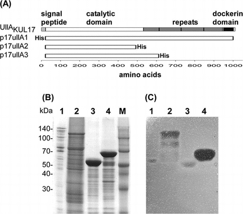
Table 1. Primers and plasmids used in this study.
Fig. 1. SDS-PAGE analysis of proteins separated by phenyl Sepharose chromatography.
Notes: Active fractions after Q Sepharose chromatographies of KUL17-secreted proteins were loaded on phenyl Sepharose column. Fractions were analyzed on 10% SDS-PAGE and stained by CBB R-250 (A) and activity staining using ulvan gel (B). Fraction numbers are shown on the top of each gel. Q indicates pooled fractions after Q Sepharose. M1 and M2 are molecular weight markers. Molecular weights are shown on the left in kDa for M1 and on the right in kDa for M2. Arrowhead indicates the candidate of ulvan-degrading enzyme.
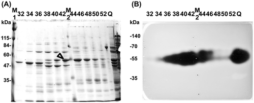
Fig. 2. Identification of ulvan-degrading enzyme.
Notes: Active fractions of secreted proteins from KUL17 after phenyl Sepharose column chromatography were pooled and analyzed on SDS-PAGE. Separated proteins were visualized with (1) CBB R-250 and (2) ulvan-gel activity staining. Protein band “a” was subjected to N-terminal amino acid sequence analysis. Molecular weight size maker was shown on the left in kDa.
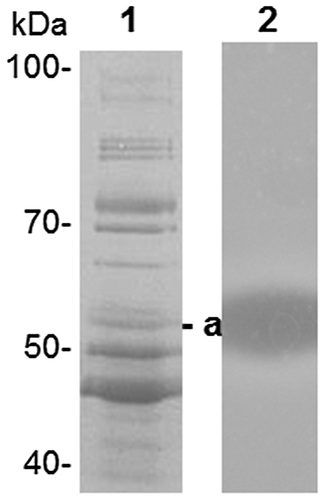
Fig. 3. Domain structure and purification of UllA.
Notes: Domain structure of UllAKUL17 is indicated in (A). Search for signal peptide, repeat sequence, and conserved domain is conducted with SignalP 4.1 Server (https://www.cbs.dtu.dk/services/SignalP/,Citation27) RADAR (https://www.ebi.ac.uk/Tools/pfa/radar/),Citation28) and Conserved Domain Search (https://www.ncbi.nlm.nih.gov/Structure/cdd/wrpsb.cgi,Citation29) respectively. Plasmids containing full length and C-terminal truncated version of UllAKUL17 are listed in (A). All plasmids are lacking signal sequences. “His” indicates the addition of His-tag sequence. (B) CBB R-250 staining of 10% SDS-PAGE gel. Lane 1; secreted protein from KUL17, lanes 2, 3, and 4; purified protein from BL21(DE3) containing p17UllA1, p17UllA2, and p17UllA3, lane M; molecular weight marker shown on the left in kDa. (C) Activity staining with ulvan-containing agarose gel. Lanes are same as in (B).

Fig. 4. Enzyme assay of UllA1.
Notes: Purified UllA1 (0.2 μg) from KUL17 was incubated with 0.4% (w/v) ulvan extracted from U. ohnoi in the buffer (20 mM Tris-HCl (pH 8.0) and 500 mM NaCl). Absorbance at 235 nm was monitored with spectrophotometer (GeneQuant 1300, GE Healthcare).
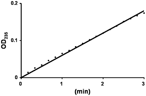
Fig. 5. Analysis of reaction products of ulvan by UllA3.
Notes: 0.4% (w/v) ulvan was mixed with 0.2 g of purified UllA3KUL17 in the buffer (20 mM Tris-HCl (pH 8.0) and 500 mM NaCl) at 0, 1, 8 and 24 h. Enzyme reaction was stopped by boiling for 10 min and filtered. The products were analyzed on Superdex Peptide 10/300 GL column at flow rate of 0. 5 ml min−1. Signal was detected by a RI detector. When 2 μg of the enzyme was used, chromatogram was indicated on the top. Ulvan showed the undigested ulvan and DP2, DP4, DP6, and DP8 were digested oligosaccharides.
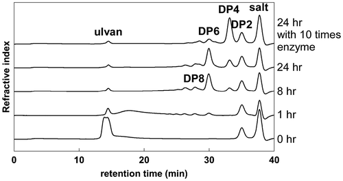
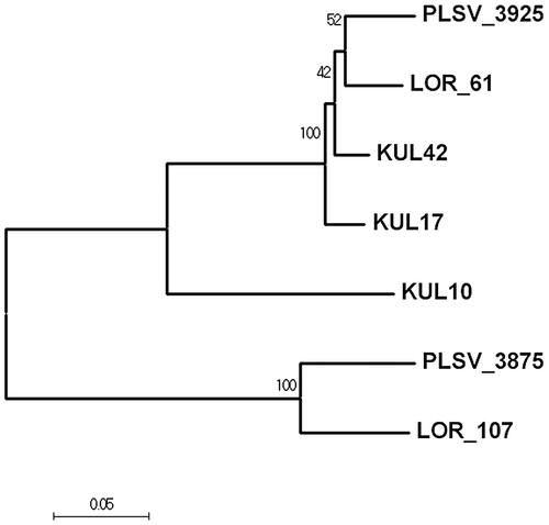
Fig. 6. Evolutionary relationship of long-type ulvan lyase. The evolutionary history was inferred using the Neighbor-Joining method.Citation30)
Notes: The percentage of replicate trees in the bootstrap test (1000 replicates) is shown next to the branches. The evolutionary distance is in the units of the number of amino acid substitutions per site. Evolutionary analyses were conducted in MEGA7.Citation31) Amino acid sequences of long-type ulvan lyase are: LOR_61; Alteromonas sp. LOR (WP_032096165.1), PLSV_3925; Alteromonas sp. PLSV (WP_033186955.1), KUL10; Glaciecola sp. KUL10 (BAY00693.1), KUL17; Alteromonas sp. KUL17 (BAY00694.1), KUL42; Alteromonas sp. KUL42 (BAY00695.1). As an outgroup, two short-type ulvan lyases, LOR_107 from Alteromonas sp. LOR (KU168251) and PLSV_3875 from Alteromonas sp. PLSV (KU168251), are included.