Figures & data

Fig. 1. Effect of TECA treatment on the viability of 2D monolayer cultured (A) and 3D spheroid cultured HDP cells (B).
Notes: Cells were treated with the indicated doses of TECA for 48 h followed by a WST-1 assay. Data are shown as mean ± SD of results from three independent experiments. Values of p < 0.05 were considered to be statistically significant. 2D MC, two-dimensional monolayer cultured; 3D SC, three-dimensional spheroid cultured.
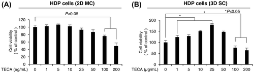
Fig. 2. Comparison of spheroid formation of control and TECA-treated HDP cells.
Notes: (A) Phase images of spheroids of control and TECA-treated HDP cells. Cells were seeded into low attachment culture plate to induce spheroids. Images were captured after 48 h of TECA treatment. The diameters of spheroids were quantified (B). Data are shown as mean ± SD of results from three independent experiments. Values of p < 0.05 were considered to be statistically significant.
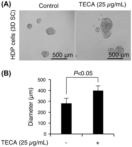
Fig. 3. Effects of TECA treatment on Wnt/β-catenin signaling activity in 3D spheroid cultured HDP cells.
Notes: (A) The luciferase activity of TCF/LEF reporter plasmids (TOPFLASH) in TECA-treated cells. HEK293 cells were co-transfected with the reporter constructs and β-galactosidase plasmids. After 48 h of TECA treatment, luciferase activity was evaluated by normalizing the levels to β-galactosidase activity. The expression levels of target genes of Wnt/β-catenin signaling, WNT5A (B) and LEF1 (C), were measured using qRT-PCR. Data are shown as mean ± SD of results from three independent experiments. Values of p < 0.05 were considered to be statistically significant.
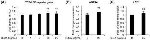
Fig. 4. Effects of TECA treatment on JAK/STAT signaling activity in 3D spheroid cultured HDP cells.
Notes: The luciferase activity of STAT5 (A) and STAT3 (C) reporter plasmids in TECA-treated cells. HEK293 cells were cotransfected with the reporter constructs and β-gal plasmids. After 48 h of TECA treatment, luciferase activity was evaluated by normalizing the levels to β-gal activity. The expression levels of SOCS1 (B) and SOCS3 (D) mRNAs were measured using qRT-PCR. Data are shown as mean ± SD of results from three independent experiments. Values of p < 0.05 were considered to be statistically significant.
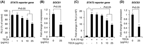
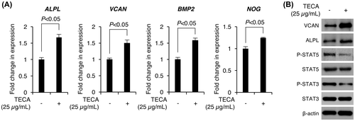
Fig. 5. Effects of TECA treatment on the expression levels of DP signature genes in 3D spheroid cultured HDP cells.
Notes: (A) The expression levels of DP signature genes including ALPL, VCAN, BMP2, and NOG were measured using qRT-PCR with their specific primers in control and TECA-treated HDP cells. Data are shown as mean ± SD of results from three independent experiments. Values of p < 0.05 were considered to be statistically significant. (B) The protein levels of DP signature genes and phosphorylated STAT proteins in TECA-treated HDP cells. HDP cells were grown in 3D cultured system followed by TECA treatment for 48 h. Cells were lysed and the indicated protein levels were examined using immunoblotting with their specific antibodies. β-actin was used as a loading control.