Figures & data
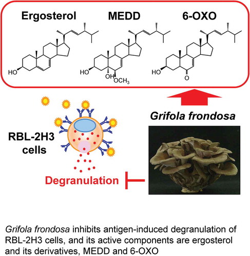
Figure 1. Effect of GFE on β-hexosaminidase release in antigen-stimulated RBL-2H3 cells.
IgE-sensitized RBL-2H3 cells pretreated with GFE or tranilast (Tra) for 20 minutes were stimulated with DNP-HSA (20 ng/mL) for 30 minutes, and the release of β-hexosaminidase was measured. Each bar represents the mean ± SEM (n = 4). *** p < 0.001 vs. Control (Cont, DNP-HSA alone).
Figure 2. Structures of active compounds and effect on β-hexosaminidase and histamine release in antigen-stimulated RBL-2H3 cells.
Structures of ergosterol (A), 6β-methoxyergosta-7,22-dien-3β,5α-diol (MEDD) (B), and 6-oxoergosta-7,22-dien-3β-ol (6-OXO) (C). IgE-sensitized RBL-2H3 cells pretreated with active compounds, GFE, or tranilast (Tra) were stimulated with DNP-HSA, and the release of β-hexosaminidase (D) and histamine (E) were measured. Each bar represents the mean ± SEM (n = 3). * p < 0.05, *** p < 0.001 vs. Control (Cont, DNP-HSA alone).
Figure 3. Effect of sterols, ergosterol, cholesterol, β-sitosterol, and campesterol on β-hexosaminidase and histamine release in RBL-2H3.
IgE-sensitized RBL-2H3 cells pretreated with 50 μM of sterol or tranilast (Tra) were stimulated with DNP-HSA, and the release of β-hexosaminidase (A) and histamine (B) were measured. Each bar represents the mean ± SEM (n = 3). * p < 0.05, *** p < 0.001 vs. Control (Cont, DNP-HSA alone). Erg, ergosterol; Cho, cholesterol; Sit, β-sitosterol; Cam, campesterol.
Figure 4. Effects of ergosterol on antigen-induced increase in IL-4 and TNF-α mRNA expression.
IgE-sensitized RBL-2H3 cells pretreated with ergosterol (Erg) were stimulated with DNP-HSA. Two hours after antigen stimulation, total RNA was extracted and IL-4 (A), TNF-α (B), and GAPDH mRNA expression was measured by quantitative RT-PCR. Values are normalized to those of GAPDH mRNA and the mean value of the non-stimulated cells (None) was set as 1.0. Each bar represents the mean ± SEM (n = 4). * p < 0.05, ** p < 0.01, *** p < 0.001 vs. Control (Cont, DNP-HSA alone).
Figure 5. Effect of ergosterol on the antigen-induced aggregation of FcεRI.
IgE-sensitized RBL-2H3 cells pretreated with ergosterol (Erg, 50 μM) or methyl beta cyclodextrin (MβCD, 1 mM) were stimulated with DNP-HSA. IgE/α-chain of FcεRI complexes were detected using anti-rat IgE antibody (A). The arrowhead shows the aggregation positive cells (A, lower panels). Data are represented as the number of aggregation positive cells/total cell number (B). Each bar represents the mean ± SEM (n = 6). *** p < 0.001 vs. Cont (Control, DNP-HSA alone).
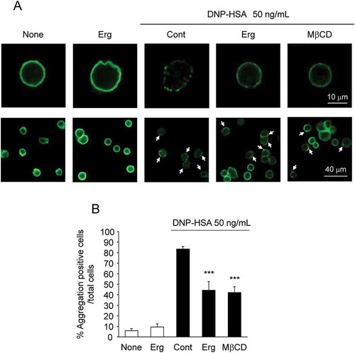
Figure 6. Effect of ergosterol on antigen-induced global tyrosine phosphorylation of proteins at 72 kDa and 75 kDa.
IgE-sensitized RBL-2H3 cells pretreated with or without ergosterol (Erg, 50 μM) were stimulated with DNP-HSA (50 ng/mL) for the indicated times. Cell lysates were blotted with anti-phosphotyrosine antibody. The arrowheads on the left side indicated the position of the molecular weight markers and the right side indicated the molecular weight of the target proteins calculated from the calibration curve obtained by the molecular weight markers.Grifola frondosa inhibits antigen-induced degranulation of RBL-2H3 cells, and its active components are ergosterol and its derivatives, MEDD and 6-OXO.