327
Views
15
CrossRef citations to date
0
Altmetric
Biochemistry & Molecular Biology

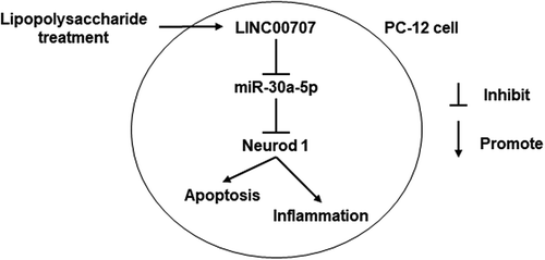
Suppression of LINC00707 alleviates lipopolysaccharide-induced inflammation and apoptosis in PC-12 cells by regulated miR-30a-5p/Neurod 1

, , , , , & show all
Pages 2049-2056 | Received 09 Apr 2019, Accepted 19 Jun 2019, Published online: 04 Jul 2019

Figures & data

Figure 1. LPS significantly inhibited proliferation and promoted apoptosis, the levels of IL-1β, IL-6, and TNF-α, and LINC00707 expression in PC-12 cells in a dose-dependent manner. (a) Proliferation was measured by MTS assay and apoptosis and cycle was measured by flow cytometry in PC-12 cells at 24 h after 0, 5, and 10 μg/mL LPS treatment. (b and c) Representative images of apoptosis and cell cycle. (d) The levels of IL-1β, IL-6, and TNF-α were measured by ELISA in PC-12 cells at 24 h after 0, 5, and 10 μg/mL LPS treatment. (e) LINC00707 expression were measured by qRT-PCR in PC-12 cells at 24 h after 0, 5, and 10 μg/mL LPS treatment. ***P < 0.001.
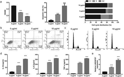
Figure 1. LPS significantly inhibited proliferation and promoted apoptosis, the levels of IL-1β, IL-6, and TNF-α, and LINC00707 expression in PC-12 cells in a dose-dependent manner. (a) Proliferation was measured by MTS assay and apoptosis and cycle was measured by flow cytometry in PC-12 cells at 24 h after 0, 5, and 10 μg/mL LPS treatment. (b and c) Representative images of apoptosis and cell cycle. (d) The levels of IL-1β, IL-6, and TNF-α were measured by ELISA in PC-12 cells at 24 h after 0, 5, and 10 μg/mL LPS treatment. (e) LINC00707 expression were measured by qRT-PCR in PC-12 cells at 24 h after 0, 5, and 10 μg/mL LPS treatment. ***P < 0.001.

Figure 2. Suppression of LINC00707 reduced inflammation, promoted proliferation, and inhibited apoptosis of LPS (5 μg/mL)-treated PC-12 cells. (a and b) LINC00707 expression was measured by qRT-PCR after transfection of siRNAs at 48 h and then treatment without or with 5 μg/mL LPS at 24 h. (c) Proliferation was measured by MTS assay. (d and e) Apoptosis and cycle was measured by flow cytometry. (f-h) The levels of IL-1β, IL-6, and TNF-α were measured by ELISA. (i and j) Representative images of apoptosis and cell cycle. ***P < 0.001.
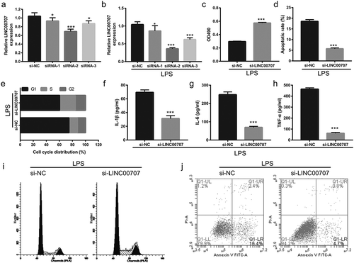
Figure 2. Suppression of LINC00707 reduced inflammation, promoted proliferation, and inhibited apoptosis of LPS (5 μg/mL)-treated PC-12 cells. (a and b) LINC00707 expression was measured by qRT-PCR after transfection of siRNAs at 48 h and then treatment without or with 5 μg/mL LPS at 24 h. (c) Proliferation was measured by MTS assay. (d and e) Apoptosis and cycle was measured by flow cytometry. (f-h) The levels of IL-1β, IL-6, and TNF-α were measured by ELISA. (i and j) Representative images of apoptosis and cell cycle. ***P < 0.001.

Figure 3. LINC00707 directly targeted miR-30a-5p in LPS-treated PC-12 cells. (a) Potential binding sites between LINC00707 and miR-30a-5p. (b) Luciferase activity ratio (Renilla/firefly) was measured in a luciferase reporter assay. (c) miR-30a-5p expression was measured by qRT-PCR in 0, 5, and 10 μg/mL LPS-treated PC-12 cells. (d) miR-30a-5p expression was measured by qRT-PCR after transfection with si-NC and si-LINC00707 in LPS (5 μg/mL)-treated PC-12 cells. ***P < 0.001.
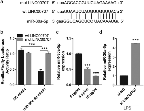
Figure 3. LINC00707 directly targeted miR-30a-5p in LPS-treated PC-12 cells. (a) Potential binding sites between LINC00707 and miR-30a-5p. (b) Luciferase activity ratio (Renilla/firefly) was measured in a luciferase reporter assay. (c) miR-30a-5p expression was measured by qRT-PCR in 0, 5, and 10 μg/mL LPS-treated PC-12 cells. (d) miR-30a-5p expression was measured by qRT-PCR after transfection with si-NC and si-LINC00707 in LPS (5 μg/mL)-treated PC-12 cells. ***P < 0.001.

Figure 4. Co-transfection of si-LINC00707 and miR-30a-5p inhibitor increased inflammation, inhibited proliferation, and promoted apoptosis in LPS (5 μg/mL)-treated PC-12 cells. (a) The expression level of miR-30a-5p was measured by qRT-PCR after only transfection of miR-30a-5p inhibitor in PC-12 cells without LPS treatment and co-transfection of si-LINC00707 and miR-30a-5p inhibitor in PC-12 cells with LPS treatment at 48 h. (b) The levels of IL-1β, IL-6, and TNF-α were measured by ELISA in PC-12 cells with LPS treatment after transfection of miR-30a-5p inhibitor. (c) The proliferation was measured by MTS in PC-12 cells with LPS treatment after transfection of miR-30a-5p inhibitor. (d) The levels of IL-1β, IL-6, and TNF-α were measured by ELISA in PC-12 cells with LPS treatment after co-transfection of si-LINC00707 and miR-30a-5p inhibitor. (e) Proliferation, and apoptosis and cycle was measured by MTS assay and flow cytometer assay, respectively, in PC-12 cells with LPS treatment after co-transfection of si-LINC00707 and miR-30a-5p inhibitor. (f and g) Representative images of apoptosis and cell cycle measured by flow cytometry. ***P < 0.001.
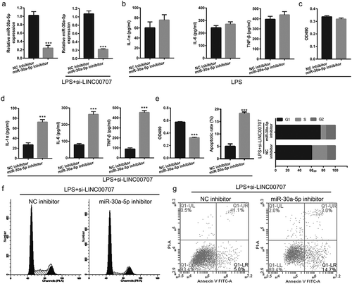
Figure 4. Co-transfection of si-LINC00707 and miR-30a-5p inhibitor increased inflammation, inhibited proliferation, and promoted apoptosis in LPS (5 μg/mL)-treated PC-12 cells. (a) The expression level of miR-30a-5p was measured by qRT-PCR after only transfection of miR-30a-5p inhibitor in PC-12 cells without LPS treatment and co-transfection of si-LINC00707 and miR-30a-5p inhibitor in PC-12 cells with LPS treatment at 48 h. (b) The levels of IL-1β, IL-6, and TNF-α were measured by ELISA in PC-12 cells with LPS treatment after transfection of miR-30a-5p inhibitor. (c) The proliferation was measured by MTS in PC-12 cells with LPS treatment after transfection of miR-30a-5p inhibitor. (d) The levels of IL-1β, IL-6, and TNF-α were measured by ELISA in PC-12 cells with LPS treatment after co-transfection of si-LINC00707 and miR-30a-5p inhibitor. (e) Proliferation, and apoptosis and cycle was measured by MTS assay and flow cytometer assay, respectively, in PC-12 cells with LPS treatment after co-transfection of si-LINC00707 and miR-30a-5p inhibitor. (f and g) Representative images of apoptosis and cell cycle measured by flow cytometry. ***P < 0.001.

Figure 5. Co-transfection of si-LINC00707 and Neurod 1-pcDNA 3.1 increased inflammation, inhibited proliferation, and promoted apoptosis in LPS (5 μg/mL)-treated PC-12 cells. (a) Neurod 1 expression was measured by western blot. (b) Neurod 1 expression was measured by qRT-PCR and western blot after transfection Neurod 1-pcDNA 3.1 at 48 h in PC-12 cells. (c) Neurod 1 expression was measured by qRT-PCR and western blot after co-transfection of si-LINC00707 and Neurod 1-pcDNA 3.1 at 48 h in LPS-treated PC-12 cells. (d) The levels of IL-1β, IL-6, and TNF-α were measured by ELISA. (e) Proliferation, and apoptosis and cycle was measured by MTS and flow cytometer assay, respectively, in PC-12 cells with LPS treatment after co-transfection of si-LINC00707 and Neurod 1-pcDNA 3.1. (f) Representative images of apoptosis. (g) Representative images of cell cycle. ***P < 0.001.
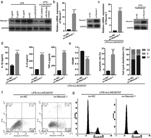
Figure 5. Co-transfection of si-LINC00707 and Neurod 1-pcDNA 3.1 increased inflammation, inhibited proliferation, and promoted apoptosis in LPS (5 μg/mL)-treated PC-12 cells. (a) Neurod 1 expression was measured by western blot. (b) Neurod 1 expression was measured by qRT-PCR and western blot after transfection Neurod 1-pcDNA 3.1 at 48 h in PC-12 cells. (c) Neurod 1 expression was measured by qRT-PCR and western blot after co-transfection of si-LINC00707 and Neurod 1-pcDNA 3.1 at 48 h in LPS-treated PC-12 cells. (d) The levels of IL-1β, IL-6, and TNF-α were measured by ELISA. (e) Proliferation, and apoptosis and cycle was measured by MTS and flow cytometer assay, respectively, in PC-12 cells with LPS treatment after co-transfection of si-LINC00707 and Neurod 1-pcDNA 3.1. (f) Representative images of apoptosis. (g) Representative images of cell cycle. ***P < 0.001.

Reprints and Corporate Permissions

Please note: Selecting permissions does not provide access to the full text of the article, please see our help page How do I view content?

To request a reprint or corporate permissions for this article, please click on the relevant link below:

Academic Permissions

Please note: Selecting permissions does not provide access to the full text of the article, please see our help page How do I view content?

Obtain permissions instantly via Rightslink by clicking on the button below:

If you are unable to obtain permissions via Rightslink, please complete and submit this Permissions form. For more information, please visit our Permissions help page.