
Figures & data
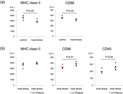
Figure 1. Administration of LC-Plasma inhibited the decrease in splenic pDC activity caused by heat stress. (a) Shown is the influence of heat stress on the expression of activation markers on pDCs from SPN. Both control and heat stress groups consisted of 4 mice. (b) Shown is the effect of the administration of LC-Plasma on the expression of activation markers on pDCs of SPN under heat stress conditions. Heat stress and heat stress + LC-Plasma groups consisted of 5 mice and 6 mice, respectively. The expression levels of MHC class II, CD86 and CD40 are shown as median fluorescence intensities (M.F.I.). Short lines represent the mean values. Statistical comparisons were performed using the Student’s t-test.
Supplemental material