
Figures & data
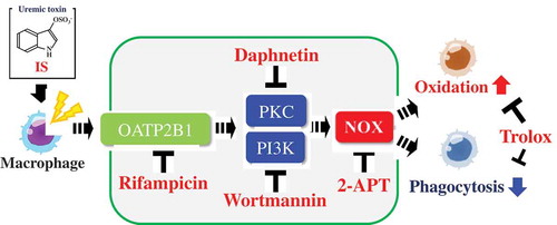
Figure 1. Effect of IS on the intracellular oxidation level in HL-60-differentiated macrophage cells (a), HL-60-differentiated granulocytic neutrophil cells (b) and undifferentiated HL-60 human leukemic cells (c). DCF fluorescence intensity was fluorometrically determined at Ex. 480 nm and Em. 530 nm. Data shown represent mean ± S.D. from four independent experiments. Using Dunnett test, significant difference from control was considered at *P< .05 or **P < .01. DCF; dichlorofluorescene, IS; indoxyl sulfate.
Figure 2. Effect of IS on the phagocytic activity of HL-60-differentiated macrophage cells. Data shown represent mean ± S.D. from four independent experiments. Phagocytic activity was fluorometrically determined at Ex. 365 nm and Em. 492 nm. Using Dunnett test, significant difference from control was considered at *P < .05 or **P < .01. IS; indoxyl sulfate.
Figure 3. Effect of 2-APT (an NADH oxidase inhibitor) (a,b), rifampicin (an OATP2B1 inhibitor) (c,d), daphnetin (a PKC inhibitor) (e,f), wortmannin (a PI3K inhibitor) (g,h) and Trolox (an antioxidant) (i,j) on the intracellular oxidation level (a,c,e,g,i) and the phagocytic activity (b,d,f,h,j) of HL-60-differentiated macrophage cells. Data shown represent mean ± S.D. from four independent experiments. Tukey–Kramer’s test was conducted for the multiple comparison and values not sharing a common superscript letter are considered significantly different at P< .05. DCF; dichlorofluorescene, 2-APT; 2-acetylphenothiazine, OATP2B1; organic anion transporter polypeptide2B1, PKC; protein kinase C, PI3K; phosphoinositide 3-kinase, IS; indoxyl sulfate.
Data availability statement
The data described in this article are openly available in the Open Science Framework at DOI:10.17605/OSF.IO/TPA6U.