Figures & data

Table 1. Coaggregation activity of LAB and M. guilliermondii in LA solution or sterile water at room temperature.
Figure 1. Effect of coincubating M. guilliermondii and their metabolites on the viability of L. pentosus and L. paracasei in LA buffer (pH 3.0) at 10°C. Values are expressed as the means of triplicate experiments (n = 3), with error bars representing the SD. Different letters indicate significant differences (p < 0.05, by Scheffe’s test) within the same day.
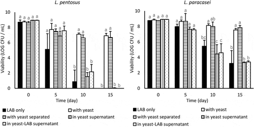
Table 2. The effect of M. guilliermondii on the LA concentration in the supernatant of LA buffer in which LAB cells were incubated at 10°C.
Figure 2. Effect of coincubating non-coaggregative yeast and MnO2 on the viability of L. pentosus and L. paracasei in LA buffer (pH 3.0) at 10°C. Values are expressed as means of triplicate experiments (n = 3), with error bars representing the SD. Different letters indicate significant differences (p < 0.05, by Scheffe’s test) within the same day.
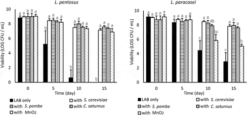
Figure 3. Effect of coincubating yeast or MnO2 on the extracellular H2O2 concentration in LA buffer in which LAB cells were incubated at 10°C. Values are expressed as means of triplicate experiments (n = 3), with error bars representing the SD. Different letters indicate significant differences (p < 0.05, by Scheffe’s test) throughout the 15-day experiment.
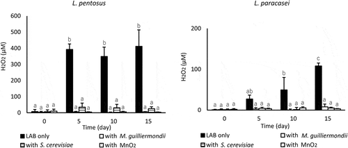
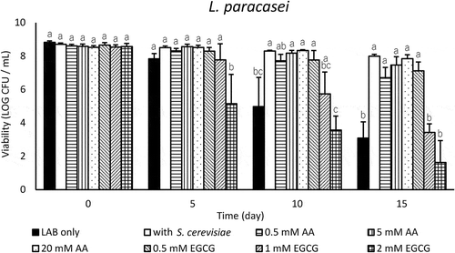
Figure 4. Effect of antioxidants on the viability of L. paracasei in LA buffer (pH 3.0) at 10°C. Values are expressed as means of triplicate experiments (n = 3), with error bars representing the SD. Different letters indicate significant differences (p < 0.05, by Scheffe’s test) within the same day.