Figures & data
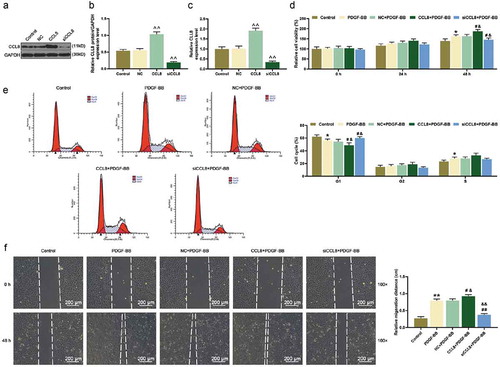
Table 1. Primers of qRT-PCR.
Figure 1. Cell viability, CCL8, and α-SMA expression in HASMCs under PDGF-BB stimulation were detected. (a) Cell Counting Kit-8 (CCK-8) assay was used to detect the cell viability after PDGF-BB stimulation at 0, 24, and 48 h. *P < 0.05, vs. Control. (b) Immunofluorescence staining was used to detect α-SMA in HASMCs stimulated by PDGF-BB at 0 and 24 h. magnification: 400 ×. (c-d) The protein expression level of α-SMA and CCL8 in HASMCs after PDGF-BB stimulation was detected by western blot. **P < 0.01, vs. Control. (e) The mRNA expression level of α-SMA and CCL8 in HASMCs after PDGF-BB stimulation was detected by qRT-PCR. **P < 0.01, vs. Control. HASMCs: human aortic smooth muscle cells; PDGF-BB: platelet-derived growth factor BB; CCL8: C-C motif Chemokine ligand 8.
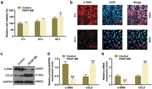
Figure 2. Effects of CCL8 on proliferation, migration, and cell cycle in HASMCs stimulated by PDGF-BB. (a-b) CCL8 protein level after upregulating or silencing CCL8 in HASMCs or PDGF-BB-induced HASMCs was measured by western blot. **P < 0.01 and ***P < 0.001, vs. Control; ##P < 0.01, vs. PDGF-BB; &&P < 0.01, vs. NC+PDGF-BB; ^^P < 0.01, vs. CCL8+ PDGF-BB; §§P < 0.01 and §§§P < 0.001, vs. siCCL8+ PDGF-BB. (c) CCL8 mRNA expression level after upregulating or silencing CCL8 in HASMCs or PDGF-BB-induced HASMCs was measured by qRT-PCR. **P < 0.01 and ***P < 0.001, vs. Control; ##P < 0.01, vs. PDGF-BB; &&P < 0.01, vs. NC+PDGF-BB; ^^P < 0.01, vs. CCL8+ PDGF-BB; §§P < 0.01 and §§§P < 0.001, vs. siCCL8+ PDGF-BB. (d) The viability of PDGF-BB-induced HASMCs after upregulating or silencing CCL8 was detected by CCK-8 assay at 0, 24, and 48 h. *P < 0.05, vs. Control; #P < 0.05, vs. PDGF-BB; &P < 0.05, vs. NC+PDGF-BB. (e) Cell cycle analysis in each group was detected using flow cytometry. *P < 0.05, vs. Control; #P < 0.05, vs. PDGF-BB; &P < 0.05, vs. NC+PDGF-BB. (f) PDGF-BB-induced HASMCs migration rates after upregulating or silencing CCL8 was measured by wound-healing assay. **P < 0.01, vs. Control; &P < 0.05 and &&P < 0.01, vs. NC+PDGF-BB; #P < 0.05, and ##P < 0.01, vs. PDGF-BB. HASMCs: human aortic smooth muscle cells; PDGF-BB: platelet-derived growth factor BB; CCL8: C-C motif Chemokine ligand 8; NC: negative control.
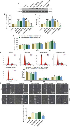
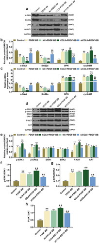
Figure 3. Effects of CCL8 on the expression of α-SMA, Sm22α, OPN, cyclinD1, p-ERK1/2, and p-AKT were detected by western blot and qRT-PCR. (a-b) Western blot assay was used to measure the protein expression level of α-SMA, Sm22α, OPN, and cyclinD1 in HASMCs stimulated by PDGF-BB after upregulating or silencing CCL8. **P < 0.01, vs. Control; #P < 0.05 and ##P < 0.01, vs. PDGF-BB; &P < 0.05 and &&P < 0.01 vs. NC+PDGF-BB. (c) The relative mRNA expressions level of α-SMA, Sm22α, OPN, and cyclinD1 in HASMCs stimulated by PDGF-BB after upregulating or silencing CCL8 was measured by qRT-PCR. **P < 0.01, vs. Control; &P < 0.05 and &&P < 0.01, vs. NC+PDGF-BB; #P < 0.05 and ##P < 0.01, vs. PDGF-BB. (d-e) Western blot assay was adopted to measure the protein expression levels of p-ERK1/2, ERK1/2, p-AKT as well as AKT in HASMCs stimulated by PDGF-BB after CCL8 upregulation or silencing. **P < 0.01, vs. Control; &P < 0.05 and &&P < 0.01, vs. NC+PDGF-BB; #P < 0.05 and ##P < 0.01, vs. PDGF-BB. (f) The phosphorylation level of ERK1 in HASMCs stimulated by PDGF-BB after upregulating or silencing CCL8 was detected by western blot. **P < 0.01, vs. Control; #P < 0.05, vs. PDGF-BB; &P < 0.05, vs. NC+PDGF-BB. (g) The phosphorylation level of ERK2 in HASMCs stimulated by PDGF-BB after upregulating or silencing CCL8 was determined by western blot. **P < 0.01, vs. Control; #P < 0.05, vs. PDGF-BB; &P < 0.05, vs. NC+PDGF-BB. (h) The phosphorylation level of AKT in HASMCs stimulated by PDGF-BB after upregulating or silencing CCL8 was quantified by western blot. **P < 0.01, vs. Control; #P < 0.05, vs. PDGF-BB; &P < 0.05, vs. NC+PDGF-BB. HASMCs: human aortic smooth muscle cells; PDGF-BB: platelet-derived growth factor BB; CCL8: C-C motif Chemokine ligand 8; NC: negative control.