Figures & data
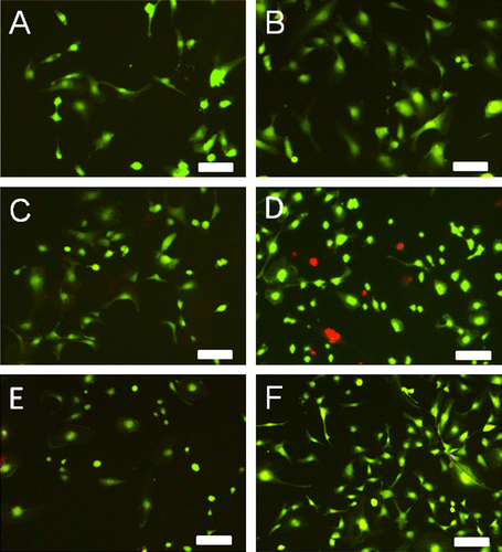
Figure 5 Digital images of HUVECs grown on cellulose–acetate-based dual-polymer electrospun scaffolds for 3 (A, C, E) or 5 (B, D, F) days. Scaffold formulations were CA-S/CA-C (A, B), Chito-S/Chito-C (C, D) or CA-S/Chito-C (E, F). Cells were stained with calcein (live, green) and ethidium (red, dead) as a live dead cell cytotoxicity assay. Scale bars are 100 μm. This figure is published in colour in the online edition of this journal that can be accessed via http://dx.doi.org/10.1080/09205063.2013.775835