Figures & data
FIGURE 1. Clinical photographs. (A) Right eye: keratic precipitates (white arrows), iris nodules (red arrow). (B) Left eye: posterior synechiae (white arrow), complicated cataract (black arrow), iris nodule (red arrow). (C) Left eye: iris nodules (red arrow), magnified image. (D) Swelling of knee joints.
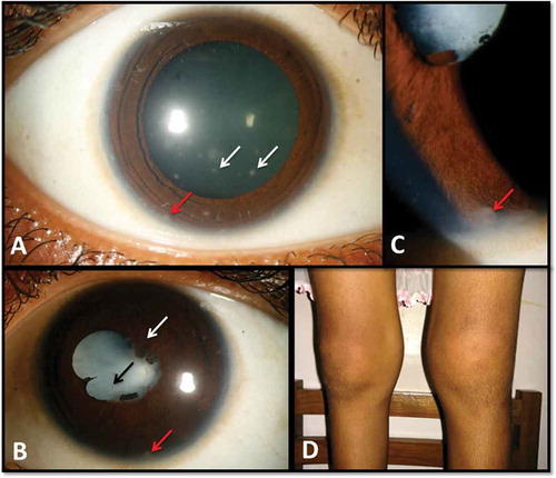
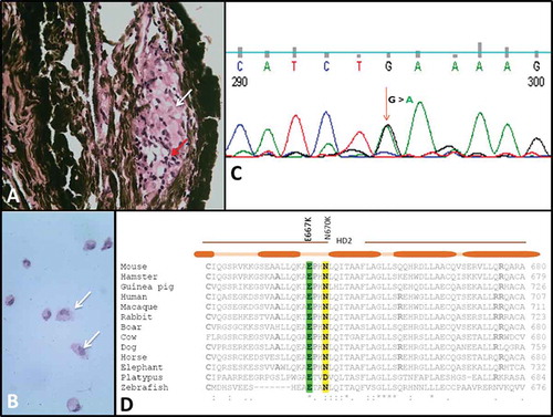
FIGURE 2. Histopathology and DNA sequencing. (A) Left eye iris nodule biopsy: non-caseating granuloma, histiocytes (white arrow), lymphocytes (red arrow). (B) Right eye vitreous: histiocytes (white arrows). (C) G>A transition in exon 4 of NOD2. (D) Multiple sequence alignment of E667 in helical domain 2 of NACHT in NOD2.