Figures & data
Figure 1. EP3 expression in normal main and accessory lacrimal glands of a 66-year-old body donor. (a–e), Immunohistochemistry photomicrograph shows strong expression of EP3 in more than 75% of acini’s nuclei and cytoplasm of main lacrimal gland (a & b) as well as accessory lacrimal gland (d & e). (c), Interlobular ducts (marked with asterisk) are devoid of any immunostaining. (f) represents the brain tissue taken as positive control (Scale bar = 60 microns).
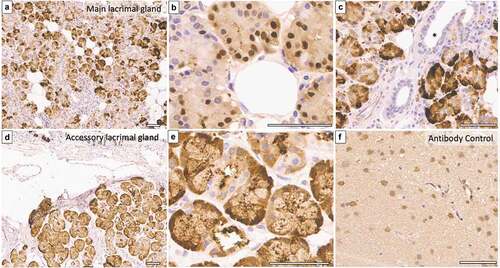
Table 1. Summary of demographics, ocular surface details and histopathology findings of normal, dacryoadenitis and SJS patients.
Figure 2. EP3 expression in main lacrimal glands of Stevens-Johnson syndrome patients. (a), Photomicrograph (h&e stained) of lacrimal gland biopsy from SJS patient (disease duration = 4 months) displays acini having atrophic changes with mononuclear cell infiltration. (b & c), EP3 receptor expression is absent within acini, only endothelial cells are expressing it weakly. (d–f), A 19-year-old SJS patient’s gland biopsy (disease duration = 48 months) show normal acinar arrangement with strong expression of EP3 and minimal inflammation. (g–i), Lacrimal gland biopsy from a 36-year-old-female shows periductular fibrosis with atrophic acini and negative EP3 immunostaining. (Scale bar = 80 microns).
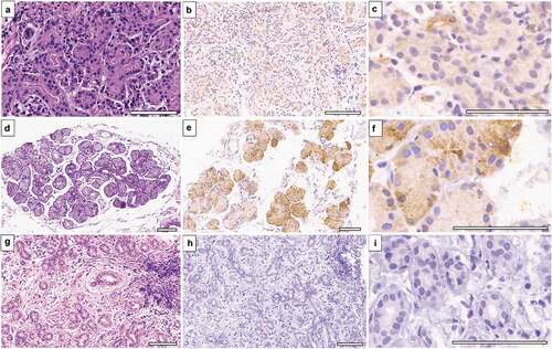
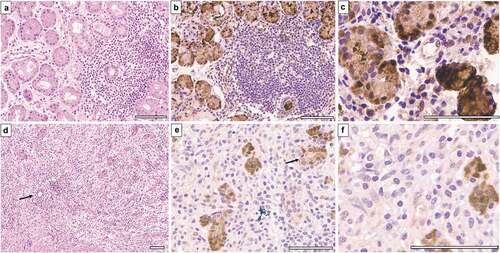
Figure 3. EP3 expression in dacryoadenitis specimens. (a) 19-year-old patient with unilateral dacryoadenitis has lymphoplasmacytic infiltrate within interstitial space surrounding normal acini. (b & c), EP3 is expressed strongly within acinar epithelium without no immunostaining in lymphocytes. (d–f), Dacryoadenitis specimen from a 17-year-old-male shows diffuse lymphoplasmacytic infiltrate with severely atrophic acini. The expression of EP3 can be observed in atrophic acini (e & f), marked with an arrow). (Scale bar = 80 microns).