Figures & data
Table 1. Baseline characteristics, management, and visual outcomes of 3 pediatric patients with CNV
Figure 1. Fundus color photography of patient 1 demonstrated parafoveal fibroglial scars on OD (A) and foveal fibroglial lesions on OS (B). Note the presence of subretinal hemorrhages (arrows) suggestive of CNV.
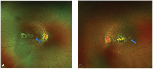
Figure 2. (A) (B) FA OD demonstrated hyperfluorescence in the early frames and late frames of CNV (yellow arrows). Note the fibroglial scars have constant hyperfluorescence. Late frame ICGA in OS (C), revealed numerous hypofluorescent spots corresponding to choroidal nodules.
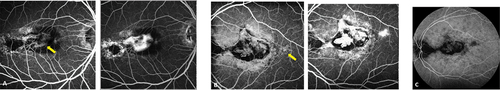
Figure 3. SD OCT (A) (B) demonstrated subretinal fibrosis, serous detachment and cystoid formations corresponding pre-epithelial choroidal neovascularization. OS (B) presence of additional macular atrophy.
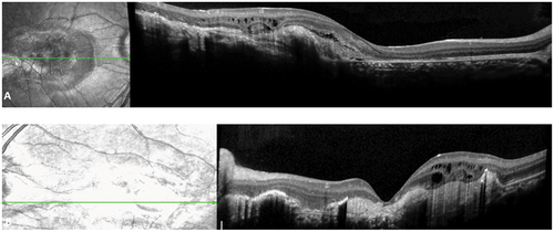
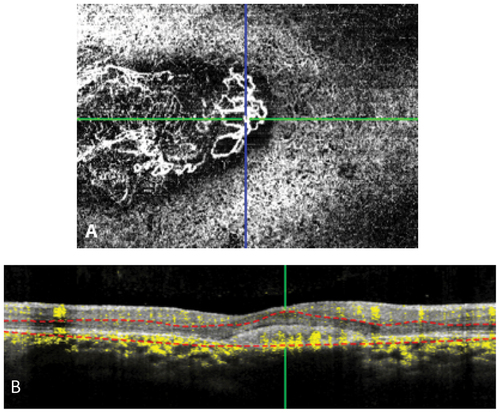
Figure 4. OCT A (A) shows CNV in the choriocapillaris layer on OS. (B) B Scan demonstrated persistent flow in CNV.