Figures & data
Table 1. Preferred host microenvironments of common pathogens in bacterial uveitis and target cells.
Figure 1. Mycobacterial infection.
Mycobacteria are taken up by phagocytes in a phagosome (a-d). Phagosomes are fused with lysosomes that provide lytic enzymes and other factors to kill the phagocytosed bacteria (e-g), the dead bacteria will then be excreted (h-j). However, peptides of degraded bacterial proteins from the lysosomes are loaded on HLA class II molecules to be presented on the surface of the phagocyte (j) and serve as antigens for T cells. Mycobacteria can either prevent the fusion of phagosomes and lysosomes (k) or even survive and proliferate in phagolysosomes. They escape from the phagolysosomes to the cytosol and multiply within their “host” cell. Degraded bacterial proteins arising during their proliferation and metabolism are now presented on HLA class I molecules on the cell surface to alert cytotoxic T cells (l). The life mycobacteria can remain latently within their host cell, abusing it as shuttle for the discrete transport within the organism (m) and finally can kill the host cell and release more mycobacteria to infect further cells (n).Citation65
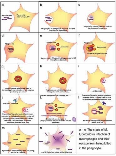
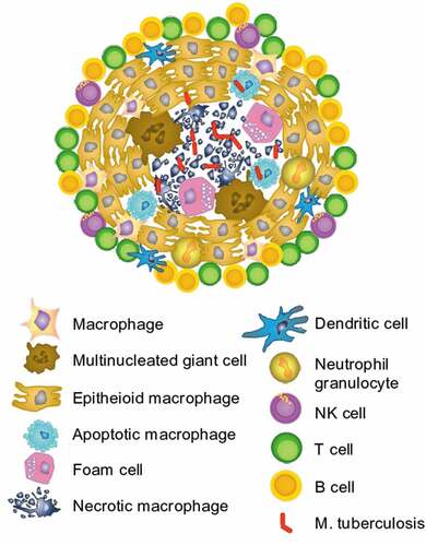
Figure 2. Granuloma in M. tuberculosis infection.
Inefficient killing of mycobacteria leads to their persistence within the host, who is nevertheless able to control the infection by forming granulomas, which enclose the mycobacteria. In that case, the macrophages are differentiating into epithelioid cells and multinucleated giant cells and form a dense nodule of cells with the mycobacteria and infected cells in the center. If the mycobacteria manage to lyse infected macrophages within the center of a granuloma, it is called “caseating” granuloma. This accumulation of differentiated macrophages with mycobacteria is surrounded by cells of the adaptive immune response like NK cells, B cells, and T cells. Those cells get respective information about the pathogen in the center, controlled by the macrophages. T cells that had been part of such granulomas are expected to be specific for mycobacterial peptides and produce IFN-γ after stimulation with mycobacterial antigens, as observed in Interferon-gamma release assays (IGRA) used as cellular in vitro-diagnosis of tuberculosis.