
Figures & data
Figure 1. Flowchart of the selection of sarcoidosis patients with an ophthalmic examination, with and without uveitis.
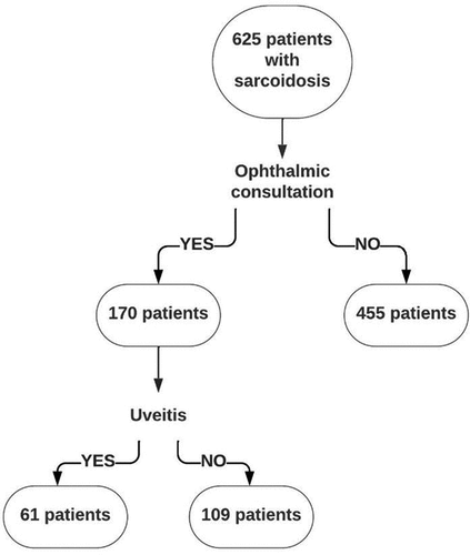
Table 1. Characteristics of the sarcoidosis patients with and without uveitis.
Table 2. Characteristics of uveitis patients with or without tumour necrosis factor (TNF) alpha inhibitor treatment.
Figure 2. Fundus images of both eyes of a 32-year-old female sarcoidosis patient with bilateral panuveitis. The black arrows indicate snowballs, the white arrows indicate multiple atrophic chorioretinal lesions.
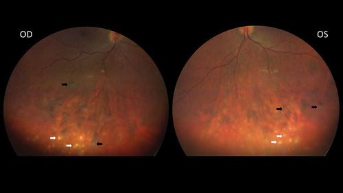
Figure 3. Fluorescein angiogram of both eyes of the same sarcoidosis patient with bilateral panuveitis. The black arrows indicate vascular leakage, the white arrows indicate hyperfluorescent dots corresponding to multiple atrophic chorioretinal lesions.
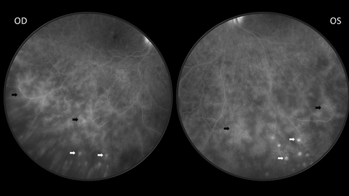
Table 3. Some relevant polymorphisms in sarcoidosis patients with and without uveitis.