Figures & data
Figure 1. Immunohistochemistry analysis expression of VEGF in the endometrium and myometrium at adenomyosis. IHC MAT to VEGF, original magnification 80×.
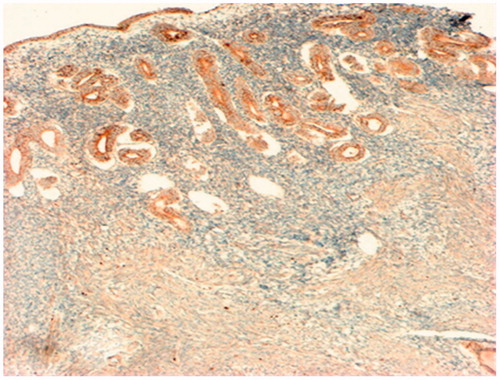
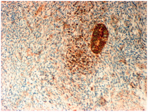
Figure 2. Immunohistochemistry analysis of VEGF Expression in the myometrium of patients with chronic pelvic pain caused by adenomyosis. A weak reaction to MAT for VEGF in the cytoplasm of smooth myocytes and intense reaction in the cells of the perivascular compartment. IHC MAT to VEGF, original magnification 200×.
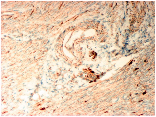
Figure 3. Immunohistochemistry analysis of VEGF Expression in the walls of venous vessels in areas of myometrium remodeling in patients with adenomyosis. IHC MAT to VEGF, original magnification 80×.
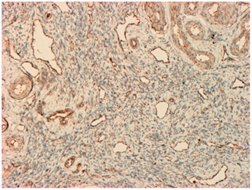
Figure 4. Immunohistochemistry analysis of VEGF Expression in foci of adenomyosis, associated mainly with the epithelial cells. IHC MAT to VEGF, original magnification 80×.
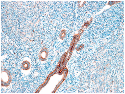
Figure 5. Immunohistochemistry analysis expression of VEGF in ectopic endometrium and surrounding myometrium stroma with signs of infiltration. IHC MAT to VEGF, original magnification 160×.