Figures & data
Figure 1. The validity of CUS to induce stress in mice and impair reproductive function. (A) Ovary weight index. (B) Mean serum CORT concentrations. (C) Number of follicles at different stages. (D) Mean cycle length of different stages of estrous cycle in the final two weeks before sacrifice. (E-J) Serum reproductive hormone concentrations. M = CUS model group; C = control group; primordial: primordial follicle; primary: primary follicle; secondary: secondary follicle; antral: antral follicle; atretic: atretic follicle; *: p < .05; **: p < .01; ***: p < .001.
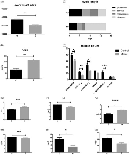
Table 1. Comparison of weekly body weight in model and control mice.
Figure 2. Protein expression analysis of FSHR and AMH in the ovaries. (A) Western blot images of FSHR and AMH in the two groups. (B) Immunoreactivity staining results showing FSHR and AMH protein expression in antral follicles and preantral follicles. Magnification: ×400. Scale bar = 12.5 μm.
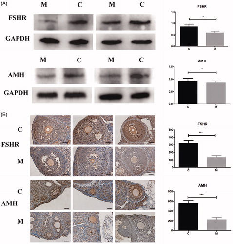
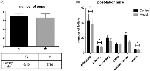
Figure 3. Fecundity of mice in both groups and long-term stress effect in mice. (A) Litter size and fertility rate of both groups. (B) The number of follicles at different stages in postlabor mice.