
Figures & data
Figure 1. Findings of virilization. (a) Hirsutism of face. (b) Male-pattern baldness (with permission from the patient).
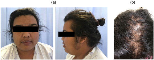
Figure 2. (a) Ultrasonographic appearance of left ovarian hyperechoic mass sized 5 × 5 cm. (b) Computed tomography of abdomen shows an enhancing lesion (7.8 × 5.2 × 4.8 cm) with contained 5.8 × 4.7 × 4.5-cm well-defined enhancing fatty mass without calcification likely arising from the left ovary (arrow).
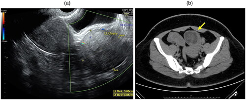
Table 1. Body weight and laboratory results before and after surgery.
Figure 3. (a) Cross-sectional appearance of the left ovary. The ovarian tumor cut surfaces are well-circumscribed, yellowish color, and lobulated. (b) Tumor cells have round to polygonal shape with eosinophilic to clear cytoplasm and oval nucleus. (c) Tumor cells arrange in cord and sheet (*) with fibrovascular tissue (arrow). (d) Tumor cells show positive for alpha-inhibin immunostaining.
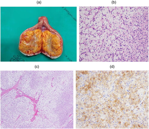
Table 2. A summary of steroid cell tumors, not otherwise specified with spontaneous pregnancy reported after surgery.