Figures & data
Figure 1. (A) Characterization of proliferative endometrial stromal cells by immunofluorescence assay for the stromal marker Vimentin (red). Scale Bar= 20 µm. (B) Representative RT-PCR analysis of vasn transcript expression in proliferative endometrial stromal cells, granulosa cells and myometrial cells of three independent cell cultures. (C) Representative western blot analysis of Vasn protein expression in proliferative endometrial stromal cells, granulosa cells and myometrial cells. Densitometric absorbance values from three independent experiments were averaged (± SEM) and were expressed as arbitrary units (a.u.). *p ≤ 0.001. ESCs = endometrial stromal cells; GCs = granulosa cells.
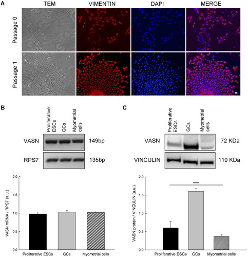
Figure 2. Localization of Vasn in primary cultures of granulosa and endometrial stromal cells by immunofluorescence staining with anti-vasorin (red) and with DAPI (Blue). Scale bar= 20 µm.
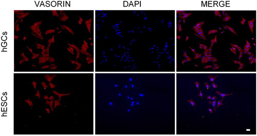
Figure 3. Vasn immunoreactivity in the human ovarian tissue. (A-B-C) Histological images stained with hematoxylin and eosin (H&E) of human ovarian tissue. Vasn expression was observed on the surface of pregranulosa cells (A’-A’’, arrowhead) and it was also maintained during follicle maturation at the surface of granulosa cells (B’-B’’, arrowhead). High Vasn expression was also present on the surface of cumulus oophorus cells surrounding the oocyte (C’-C’’, arrowhead). Scale bar= 200 µm.
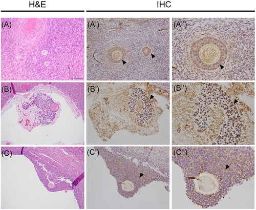
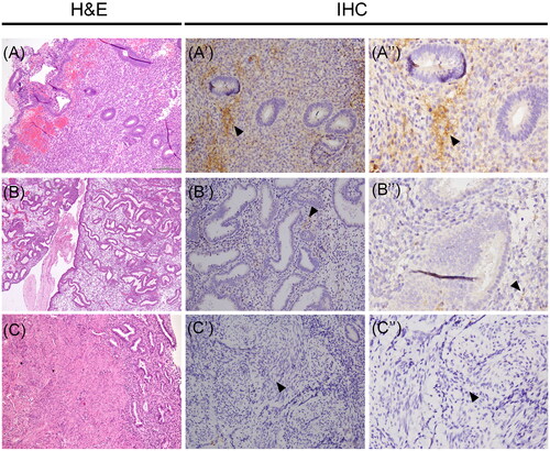
Figure 4. Vasn immunoreactivity in the normal proliferative-secretory phase endometrial tissue and normal myometrium. (A-B-C) Histological images stained with hematoxylin and eosin (H&E) of human uterine tissues. Vasn expression was evident in the stromal cells of proliferative endometrium (A’-A’’, arrow). Vasn expression was extremely weak in the stroma of secretory endometrium (B’-B’’, arrow). Vasn expression was absent in the myometrium (C’-C’’). Scale bar = 200 µm.