
Figures & data
Figure 1. mRNA expression of adhesion molecules, cytokines, and chemokines in human umbilical vein endothelial cells (HUVECs) treated with progestogens. Steroid-deprived and serum-starved HUVECs were treated for 48 h in the presence of various progestogens, namely 100 nM natural progesterone (P4), 100 nM dienogest (DNG), and 100 nM medroxyprogesterone acetate (MPA). The cells were stimulated with interleukin (IL)-1β (40 U/mL) for 4 h. Relative mRNA expression of (A) ICAM-1, (B) E-selectin, (C) IL-6, and (D) MCP-1 was measured by real-time PCR and compared with vehicle-only treatment. Data are presented as the mean ± SEM of three biological replicates. *P < 0.05 vs. vehicle only.
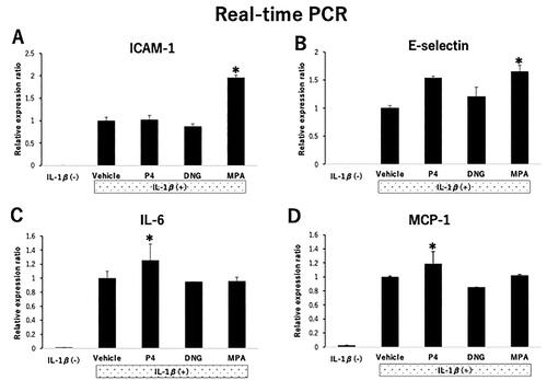
Figure 2. Effects of various progestogens on the adhesion of U937 monocytoid cells to human umbilical vein endothelial cells (HUVECs) under flow conditions.HUVECs were treated with various progestogens as described in . (A) Representative micrographs of HUVECs in a flow chamber system. The indicated small round cells were adherent to the U937 monocytoid cells. Sample fields are at 40× magnification (left), with details of the cells (right) shown at 100× magnification. (B) U937 monocytoid cells at 10,000/mL were perfused over HUVEC monolayers, and adherent cells were counted 5 min after perfusion. The total number of adherent cells in 10 randomly selected microscopic fields of each sample is shown. Data are presented as the mean ± SEM of three biological replicates. *P < 0.05 vs. vehicle only.
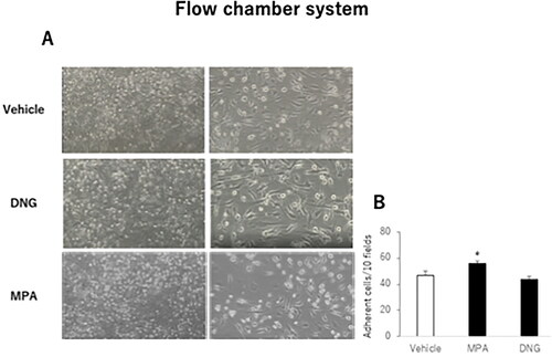
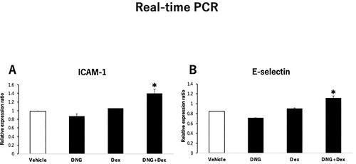
Figure 3. Effects of the GR agonist on the expression of endothelial adhesion molecules in dienogest (DNG)-treated human umbilical vein endothelial cells (HUVECs).HUVECs were treated with 100 nM DNG in the presence or absence of 100 nM dexamethasone (DEX), as described in . Relative mRNA expression of (A) E-selectin and (B) ICAM-1 in HUVECs. Data are presented as the mean ± SEM of three biological replicates.
Supplemental Material
Download MS Word (1.9 MB)Data availability statement
All data generated or analyzed during this study are included in this published article and available from the corresponding author on reasonable request.