
Figures & data
Figure 1. The Time frame of the experiments. The animals were divided into seven groups containing six rats in each group.
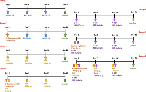
Figure 2. (a) Number of follicles. Primordial, primary, secondary, antral, and atretic follicle counts of ovaries in the all groups. ++ p < .01, +++ p < .001 ++++ p < .0001, compared to group 1; * p < .05, ** p < .01, *** p < .001, **** p < .0001, compared to group 2.2. (b) Serum concentrations of AMH.
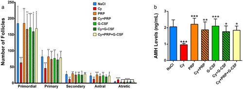
Figure 3. (a) Primary, secondary, and antral AMH positive staining follicle counts in the all groups. ++ p < .01, +++ p < .001, ++++ p < .0001, compared to group 1; * p < .05, ** p < .01, *** p < .001, **** p < .0001, compared to group 2. (b) Primary, secondary and antral AMH positive staining follicle intensity score in the all groups. ++++ p < .0001, compared to group 1; **** p < .0001, compared to group 2. 3c) Immunohistochemistry of AMH in the ovary. In the upper panel, AMH positive stained primary follicules are seen in the ovary sections. Densely stained primary follicles were observed in NaCl, PRP and G-CSF groups. In Cy group, primary follicles were stained weakly. Intensity of AMH was increased and stained moderately in Cy + PRP, Cy + G-CSF and Cy + PRP + G-CSF groups. (Bar: 50 μm) In the Middle panel, AMH positive stained secondary follicules are seen in the ovary sections. Intensity of AMH was stained strongly in PRP and G-CSF groups. In Cy group, secondary follicles were stained weakly. In NaCl, Cy + PRP, Cy + G-CSF and Cy + PRP + G-CSF groups, intensity of AMH was stained moderately. (Bar: 100 μm) In the bottom panel, AMH positive stained antral follicules are seen in the ovary sections. Intensity of AMH was stained strongly in PRP and G-CSF groups. In Cy group, secondary follicles were stained weakly. In NaCl, Cy + PRP, Cy + G-CSF and Cy + PRP + G-CSF groups, intensity of AMH was stained moderately. (Bar: 200 μm).
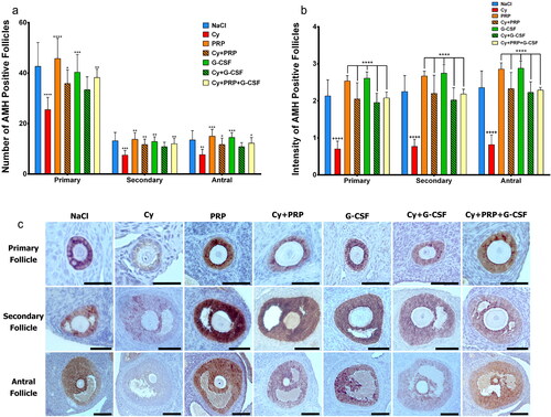
Table 1. Comparison of the primary, secondary and antral AMH-positive staining follicle counts, AMH-positive staining follicle intensity, antral INSL3 positive staining follicle intensity and primordial, primary, secondary, antral, atretic follicle counts of all groups.
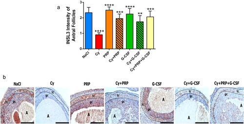
Figure 4. (a) INSL3 intensity of antral follicles. ++++ p < .0001, compared to group 1; ** p < .01, *** p < .001, **** p < .0001, compared to group 2. (b) Immunohistochemical analysis of INSL3 in the antral follicles of ovaries. Immunoreactivity of INSL3 was detected as brown staining. Strong INSL3 staining was observed in the follicular theca and granulosa cell layer in NaCl, PRP and G-CSF groups. Weakly INSL3 staining was detected both follicular theca and granulosa cell layer in Cy group. Moderate INSL3 staining was observed in Cy + PRP, Cy + G-CSF and Cy + PRP + G-CSF groups. A, antrum of antral follicle; tc, follicular theca cell layer; gc, granulosa cell layer. All figures include same magnification. Bar: 200 μm.
Data availability statement
The data sets used and/or analyzed during the current study are available from the corresponding author on reasonable request.