Figures & data
Table I. Characteristics of study population.
Figure 1. Timing of haemostatic tests and plasma concentration of C-reactive protein (CRP). Box plots of the (A) time interval between platelet reactivity and haemostatic test and positive blood culture (left panel) or start of antibiotics (right panel). (B) CRP levels at the day (+/- 24 hrs) of the haemostasis assays. Presented data are medians with IQR, minimum and maximum values. NS, not statistically significant.
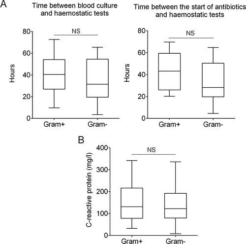
Figure 2. Platelet reactivity and platelet-monocyte complex in patients with Gram-positive and Gram-negative sepsis. (A) Platelet membrane expression of P-selectin and platelet-fibrinogen binding is depicted as median fluorescence intensity (MFI) in arbitrary units, at baseline and after stimulation with two concentrations of the platelet agonists adenosine diphosphate (ADP) and collagen-related peptide (CRP-XL) in healthy controls (n = 20), patients with Gram-positive (Gram+, n = 15) and Gram-negative (Gram-, n = 17) sepsis. (B) Platelet-monocyte complex (PMC) formation is depicted as the MFI of the platelet marker CD61 on CD14-positive cells. Data depicted are medians with IQR (platelet reactivity) or median with IQR, minimum and maximum values (PMC). * Gram-positive vs. Gram-negative, #Gram-positive vs. healthy controls.
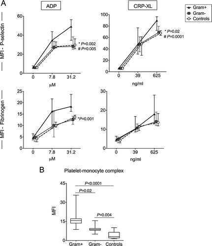
Figure 3. Plasma concentrations of platelet and coagulation activation markers. Data depicted are medians with IQR, minimum and maximum values.
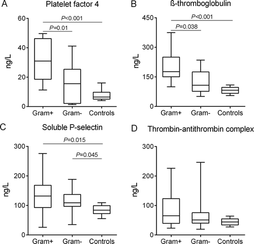
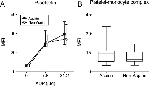
Figure 4. Platelet reactivity in aspirin and non-aspirin using patients. Data depicted are medians with IQR, minimum and maximum values.