Figures & data
Figure 1. Peripheral monocytes in active ITP patients. Peripheral monocyte percentage (a) and count (b) from healthy controls (n = 6) and active ITP patients (n = 20). The correlations of peripheral monocyte percentage (c) and count (d) with platelet count in ITP patients. Bars represent mean ± SEM. *p < 0.05; unpaired Student’s t test and Pearson’s correlation test.
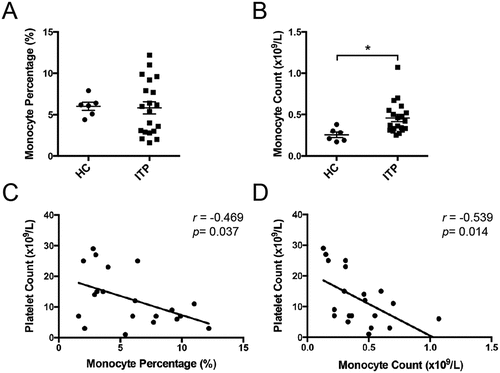
Figure 2. Plasma IFN-γ/IL-4 levels and the correlations with peripheral monocyte count in active ITP patients. Plasma IFN-γ (a) and IL-4 (b) levels from healthy controls (n = 6) and active ITP patients (n = 16). The correlations of plasma IFN-γ (c), IL-4 (d) levels and IFN-γ/IL-4 ratio (e) with peripheral monocyte count in ITP patients. Bars represent mean ± SEM. *p < 0.05; ** p < 0.01; unpaired Student’s t test and Pearson’s correlation test.

Figure 3. Changes of peripheral monocyte dynamics and plasma IFN-γ/IL-4 levels in ITP patients after eltrombopag treatment in vivo. Peripheral monocyte percentage (a), monocyte count (b), platelet count (c), plasma IFN-γ (d) and IL-4 (e) level in ITP patients (n = 11) before or after eltrombopag treatment in vivo. (f) Plasma IFN-γ/IL-4 ratio from healthy controls (n = 6) and ITP patients (n = 11) before or after eltrombopag treatment. (g) the correlations of plasma IFN-γ/IL-4 ratio (E) with peripheral monocyte count in ITP patients after eltrombopag treatment in vivo. Bars represent mean ± SEM. *p < 0.05; unpaired Student’s t test, one-way ANOVA tests and Pearson’s correlation test.
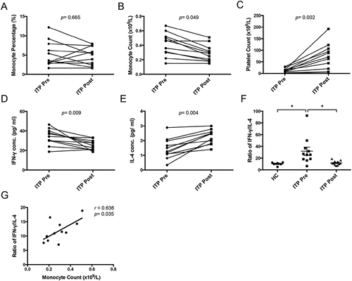
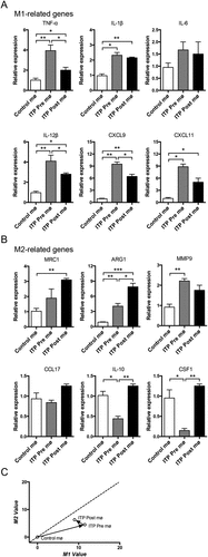
Figure 4. The activation phenotypes of ITP macrophages before or after eltrombopag treatment in vivo. Expression of M1 (a) and M2 (b) phenotype-related genes in ITP macrophages before or after eltrombopag treatment was detected by real-time PCR. For each gene, the RQ value of control macrophages from healthy individuals was designated 1.000, respectively. (c) a two-dimensional illustration of the macrophage phenotype is shown. For each gene, the value of the control macrophages from healthy individuals was designated 0. The mean values of the relative expression of genes mentioned above were used for the calculation and the M1 and M2 values. The phenotypic evolution of ITP macrophages before or after eltrombopag treatment is indicated by arrows. The results are from three independent experiments. Bars represent mean ± SEM. *p < 0.05; ** p < 0.01; *** p < 0.001; one-way ANOVA tests.