Figures & data
Table I. Centrifugation protocols according to the low speed centrifugation concept (LSCC). In all experimental set-ups, the high (710 ×g) and the low (44 ×g) centrifugation protocols were selected for comparison.
Figure 1. A. Analysis of the morphology of monocultured pOB with and without PRF treatment after 2 days. POBs were stained for SMA (green) and DAPI (blue) by immunofluorescence staining. B. Isolation of RNA from monocultured pOBs treated with PRF compared to untreated pOBs after 2 days. C. Effect of indirectly applied PRF on the viability of pOBs seeded in monoculture after 3, 7 and 9 days. Statistical significance was assessed by ****p < 0.0001. Scale bars: 500 μm.
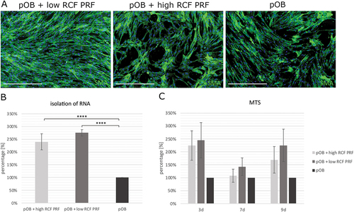
Figure 2. Determination of the relative gene expression of osteogenic differentiation factors in cultures of pOBs with and without indirect PRF treatment using specific primers for alkaline phosphatase (ALP), collagen type 1 (col-1), osteonectin (SPARC), osteopontin (SPP1), BMP-1 and BMP-2 by real time PCR after a 2-day incubation period.
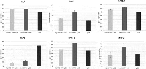
Figure 3. Evaluation of the release of the proosteogenic growth factors TGF-β1 and PDGF-BB from cell culture supernatants of monocultured pOB after 3, 7 and 9 days. Statistical significance was calculated using ANOVA and was assessed by **p < 0.01, ***p < 0.001 and ****p < 0.0001.
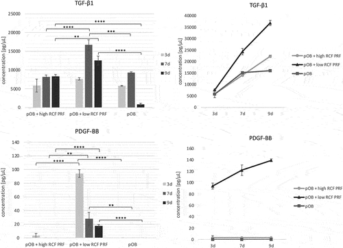
Figure 4. Analysis of microvessel-like structure formation in an in vitro co-culture model for bone tissue engineering. A. Immunofluorescence staining for the endothelial cell type specific marker CD31 of co-cultures consisting of HDMEC and pOB. Co-cultures were either treated with PRF or without PRF (control group) for 7 (upper row) and 14 (lower row) days. B. Evaluation of the relative amount of vascular structure formation after 7 and 14 days. Statistical significance was assessed by **p < 0.01 and ****p < 0.0001. Scale bars 150 μm.
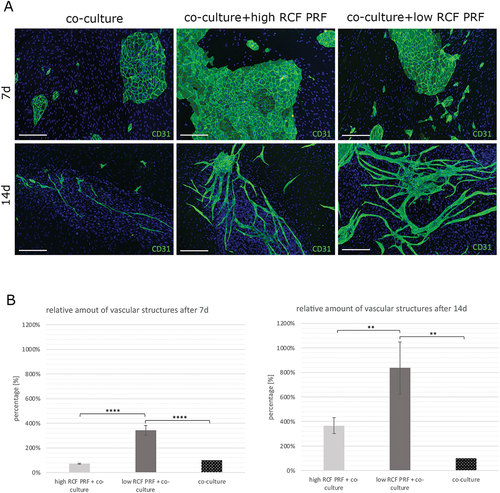
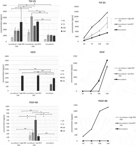
Figure 5. Evaluation of the release of the proangiogenic growth factors TGF-β1, VEGF and PDGF-BB from cell culture supernatants of co-cultures consisting of pOB and HDMEC after 4, 7, 10 and 14 days. Statistical significance was calculated using ANOVA and was assessed by *p < 0.05, **p < 0.01.