
Figures & data
Figure 1. Platelets in PRP cultured at 37°C show “spores” and increase in number.
A, PRP at different time points (6 h, 12 h, or 24 h) was cultured at 37°C and fixed with 4% paraformaldehyde. Platelets were immunofluorescence stained with phalloidin dye. B, The number of “spore” platelets at different time points (6 h, 12 h, or 24 h) was counted and analyzed. C–E, PRP at different time points (24 h or 48 h) was cultured, and then PRP was given a certain shear force. The number of platelets at different time points was counted and analyzed. Data are expressed as mean ± SD of three independent experiments.*p < .05, **p < .01, ***p < .001, ****p < .0001. Scale bars represent 5 μm.
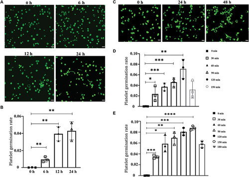
Figure 2. Different components in M199 medium promote platelet sprouting.
A, Washed human platelets were treated with M199, AMP, or MTB and cultured at 37°C for different times (6 h, 12 h, 24 h, or 36 h). B,Washed human platelets, which were treated with different components in M199 medium, was cultured at 37°C. The number of “spore” platelets at different time points (6 h, 12 h, 24 h, or 36 h) was counted and analyzed. Scale bars represent 10 μm. C, D, ΔΨm depolarization and PS externalization of platelets were treated with M199, AMP, or MTB at different time points. Data are expressed as mean ± SD of three independent experiments.
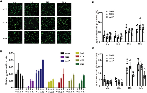
Figure 3. Activation of AMPK promotes sporulation of platelets.
A–D, PRP at different time points(0 h, 24 h, or 48 h) was cultured at 37°C, and then the expression levels of VASP,p-VASP, AMPK,p-AMPK, CAMKK in platelets were detected by Western blotting (A). And densitometry of immunoblot for p-AMPK, CAMKK, and p-VASP from the Western blot data (B–D). E, F, Washed human platelets were treated with AMPK activator AI (200 μM, 400 μM), DMSO as solvent. The number of “spore” platelets at different concentrations (0 μM, 200 μM, 400 μM) was counted and analyzed. G-I, Washed human platelets which were cultured at 37°C were treated with PKA activator Forsklin (5 μM), at different time points (0 h, 24 h, or 48 h).DMSO as solvent. The expression levels of VASP,p-VASP, AMPK, and p-AMPK, in platelets were detected by Western blotting (G). J, K, The number of “spore” platelets at different time points (0 h, 24 h, or 48 h) was counted and analyzed.Data are expressed as mean ± SD of three independent experiments.*p < .05, **p < .01, ***p < .001, ****p < .0001. Scale bars represent 5 μm.
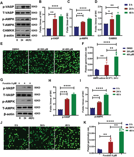
Figure 4. The cytoskeleton protein of spore-like platelets cultured in PRP has significantly a different expression.
A, B, PRP at different time points (0 h, 24 h or 48 h) was cultured at 37°C, then the expression levels of PLS1 in platelets were detected by Western blotting (A). And densitometry of immunoblot for PLS1 from the Western blot data (B). C–H,The expression levels of PLS1 and its related genes, KRT19, ESPN, CNN3, CAPZB, and DBNL were detected by PCR. I, STRING database analysis of proteins associated with PLS1.Data are expressed as mean ± SD of three independent experiments.*p < .05, **p < .01, ***p < .001, ****p < .0001.
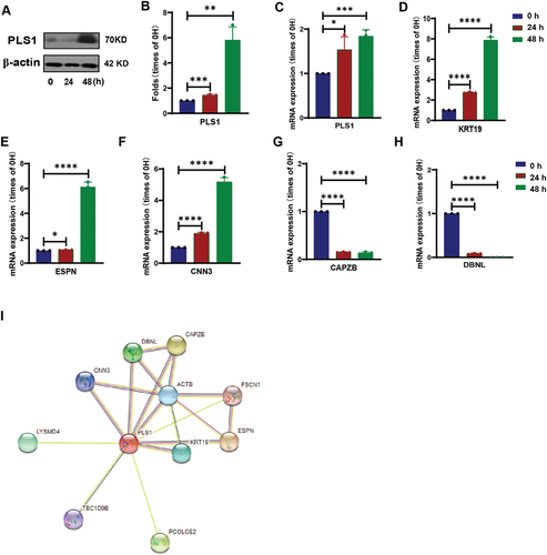
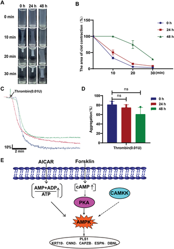
Figure 5. Spore platelets cultured in PRP still have platelet function.
A–D, PRP at different time points (0 h, 24 h, or 48 h) was cultured at 37°C, enriched platelets were obtained after washing. Washed platelets were treated with 0.01 U thrombin and placed at 37°C for different time points (0 min, 10 min, 20 min, or 30 min). The area ratio of clot contraction (B) and platelet aggregation rate (D) were analyzed. Mechanism map of platelet budding in PRP cultured at 37°C (E). Data are expressed as mean ± SD of three independent experiments. NS represents no statistical difference.
Supplemental material