
Figures & data
Figure 1. Generation of Syk S291A knock-in mice. (A) Restriction enzyme digest of PCR products from Syk S291A homozygous knock-in (SykS291A/S291A), Syk S291A heterozygous (SykS291A/WT), and WT littermate control (SykWT/WT) DNA. The oligonucleotides used for PCR are forward primer 5’- CATGCAGGAAATCTCCCTGGG and reverse primer 5’-TGTAAAGGGCAAGGCAACGTGG. Mutation of S291 to Ala, creates a site for MluI. WT PCR product is 362 bp while that of knock in is 213 and 149 bp. (B) Sequence analysis of PCR products of wild-type (WT) and S291A homozygous mouse DNA. (C) Repre-sentative Western blot (using 8% SDS-PAGE) showing that Syk S291 phosphorylation is present in CRP-stimulated WT platelets but absent in Syk S91A knock-in platelets. UN = Unstimulated sample.
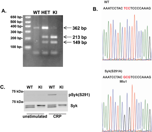
Table I. Blood cell counts from Syk S291A and WT littermate control mice. WBC = white blood cell, NE = neutrophil, LY = lymphocyte, RBC = red blood cell, PLT = platelet, MpV = mean platelet volume. *p < .05 compared to WT, n = 12.
Figure 2. Analysis of Syk isoforms in multiple tissues of Syk S291A knock-in mouse: A) Cellular extracts from different tissues from Syk S291A mice (as indicated) and wild-type littermates were run on 8% SDS-PAGE and analyzed by western blot using total Syk antibody. Two isoforms are seen in wild-type tissue extracts but only long form of Syk is seen in the tissue extracts from the Syk S291A knock in mice. B) RNA was isolated from different tissues from Syk S291A mice and wild-type litter mates was analyzed by RTPCR. The primers used were 5’-CTGAAGGAGAACCTCATCAGGG-3’ (corresponding to 841–863) as sense primer and 5’- GGCTCCTGTCCAGGTAGACC-3’ (corresponding to 1463–1442) as the antisense primer.
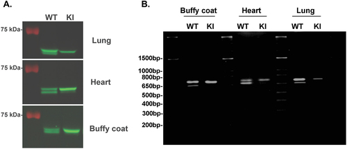
Figure 3. GPCR- and ADP-mediated platelet reactivity is intact in Syk S291A mice. (A) Representative aggregation and secretion tracings of platelets from WT and Syk S291A mice activated with the PAR4 agonist AYPGKF. (B) Quantification of ATP secretion of platelets from Syk S291A (red bars) and WT littermate control mice (green bars) stimulated with various concentrations AYPGKF. (C) Representative aggregation and secretion tracings of platelets from WT and Syk S291A mice stimulated with the P2Y agonist 2-MeSADP. (D) Quantitation of ATP secretion of platelets from Syk S291A (red bars) and WT mice (green bars) stimulated with various concentrations of 2-MeSADP. n = 3.
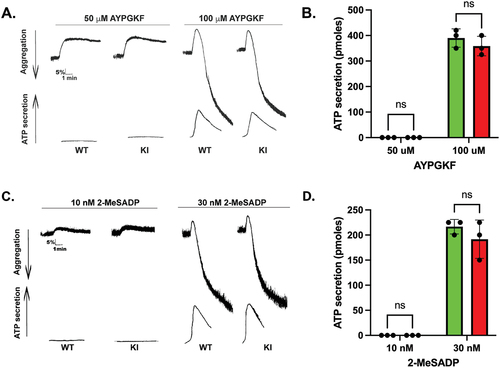
Figure 4. GPVI-mediated aggregation and secretion are potentiated in Syk S291A platelets. (A) Representative aggregation and secretion tracings of Syk S291A (red bars) and WT littermate control (green bars) platelets stimulated with the indicated concentrations of CRP, under conditions of aggregation in the absence of any feedback inhibitors. (B) Quantification of aggregation, (C) ATP secretion and (D) p-Selectin surface expression from multiple independent experiments. p. value ****<0.0001, n = 5.
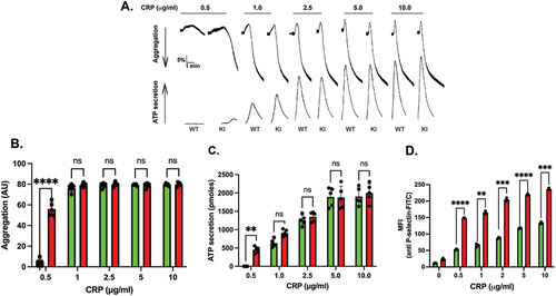
Figure 5. CRP-mediated signaling is increased in Syk S291A platelets. (A) Representative Western blots showing the indicated that phosphorylated and total protein in Syk S291A and WT littermate control platelets stimulated with the indicated concentrations of CRP for 3 minutes under aggregating conditions. (B-H) Quantification of the indicated phosphorylated residue expressed as a percentage of the maximum WT (WT10). p value****<0.0001, n = 5.
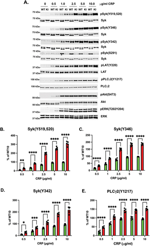
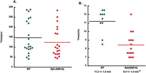
Figure 6. In vivo thrombus formation is enhanced in Syk S291A knock-in mice. (A) Scatter plot showing the time it took for bleeding to stop during tail bleeding experiments conducted on homozygous SykS291A/S291A knock-in and WT (SykWT/WT) littermate control mice 4–6 weeks of age in a blind fashion. (B) Scatter plot of the time to occlusion in WT and Syk S291A knock-in mice in a blind fashion following 7.5% FeCl3 injury on the carotid artery and analyzed by student’s test.
Supplemental Material
Download PDF (77.3 KB)Data availability statement
All data concerning this report are available in the manuscript.