218
Views
1
CrossRef citations to date
0
Altmetric
Original Articles

Variable binding by synaptic strength change

Pages 327-357 | Received 11 Sep 2008, Published online: 17 Nov 2009

Figures & data

Figure 1. Idealised binding with bind node: initial binding of red and square is later replaced by blue binding with circle.
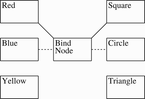
Figure 1. Idealised binding with bind node: initial binding of red and square is later replaced by blue binding with circle.

Figure 2. Sample neural firing pattern for red-square and blue-circle.

Figure 2. Sample neural firing pattern for red-square and blue-circle.

Figure 3. Topology of intra-subnet connections in the compensatory LTP binding simulation: each neuron in the base subnets connect to the bind subnet, and each neuron in the bind subnet connects to the base subnets.

Figure 3. Topology of intra-subnet connections in the compensatory LTP binding simulation: each neuron in the base subnets connect to the bind subnet, and each neuron in the bind subnet connects to the base subnets.

Table 1. Network constants.

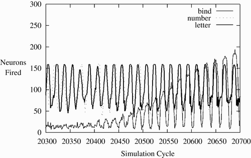
Figure 4. Neurons firing per cycle indicating CA formation.

Figure 4. Neurons firing per cycle indicating CA formation.

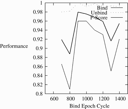
Figure 5. Effect of binding period on binding.

Figure 5. Effect of binding period on binding.

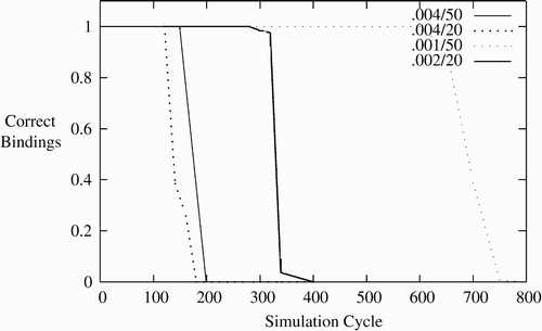
Figure 6. Duration of bindings via STP varying by reduction rate and time to bind.

Figure 6. Duration of bindings via STP varying by reduction rate and time to bind.

Figure 7. Gross topology of the simulation of binding with frames. The rule subnet inhibits the slots of the frames that are not active, and the slots are bound to the appropriate verbs and nouns via STP.
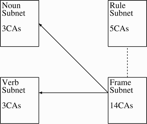
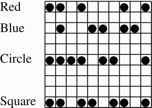
Figure 7. Gross topology of the simulation of binding with frames. The rule subnet inhibits the slots of the frames that are not active, and the slots are bound to the appropriate verbs and nouns via STP.
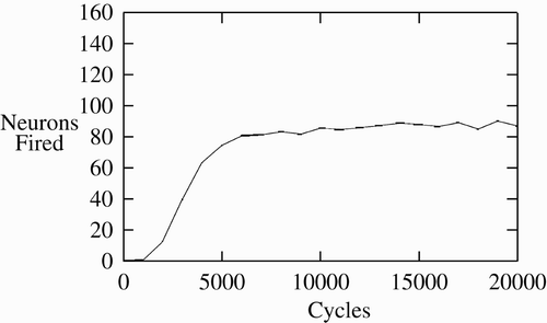
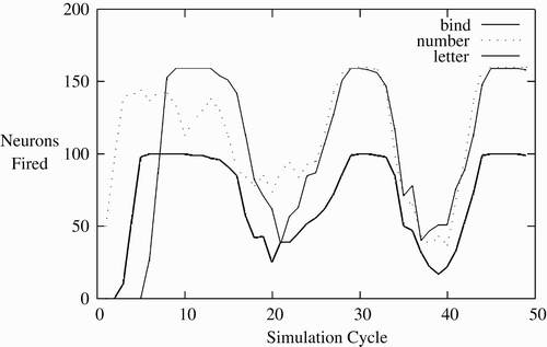
Figure 8. Firing of neurons showing synchronisation while binding.

Figure 8. Firing of neurons showing synchronisation while binding.

Figure 9. Firing of neurons showing synchronisation of items bound by STP.

Figure 9. Firing of neurons showing synchronisation of items bound by STP.

Table 2. Binding property values by method.

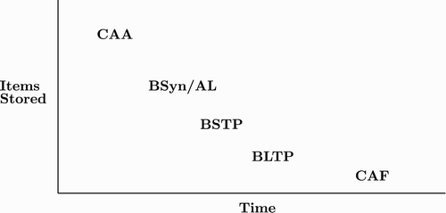
Figure 10. Memory hierarchy: different binding mechanisms provide a possible answer for the wide range of memory duration CAA, CA activation; BSyn, binding via synchrony; AL, binding via active links; BSTP, binding by STP; BLTP, binding by LTP; CAF, CA formation.

Figure 10. Memory hierarchy: different binding mechanisms provide a possible answer for the wide range of memory duration CAA, CA activation; BSyn, binding via synchrony; AL, binding via active links; BSTP, binding by STP; BLTP, binding by LTP; CAF, CA formation.

Reprints and Corporate Permissions

Please note: Selecting permissions does not provide access to the full text of the article, please see our help page How do I view content?

To request a reprint or corporate permissions for this article, please click on the relevant link below:

Academic Permissions

Please note: Selecting permissions does not provide access to the full text of the article, please see our help page How do I view content?

Obtain permissions instantly via Rightslink by clicking on the button below:

If you are unable to obtain permissions via Rightslink, please complete and submit this Permissions form. For more information, please visit our Permissions help page.