Figures & data
Figure 1. Chemical structure of neomycin.
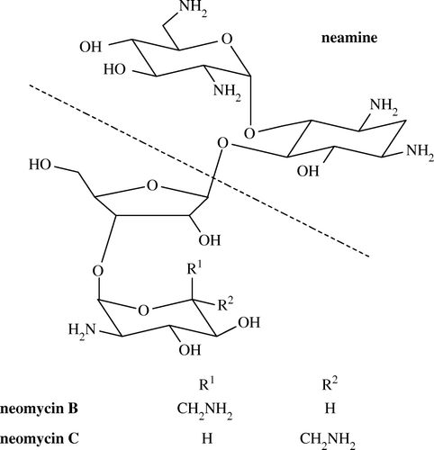
Figure 2. Titer (upper panel) and IC50 (lower panel) change curves of antisera from various bleeds of rabbits. Neomycin-OVAE was used as coating antigen at the concentration of 1 µg/ml. The titer of antiserum was defined as the dilution factor which gave 2.1 times OD values than negative serum. Each point represents the average of three well replicates.
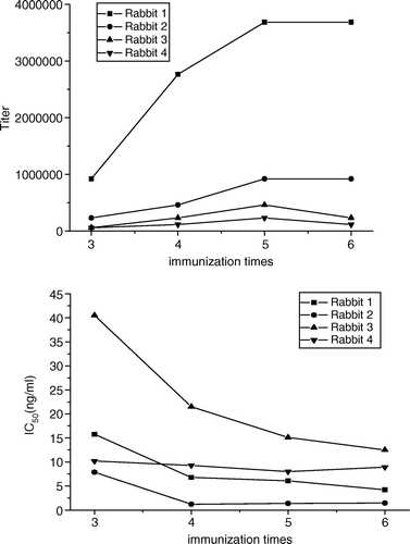
Table I. Titers and IC50 values of final antisera to neomycin-OVAE and neomycin-G-OVA.
Figure 3. Representative standard curves of the neomycin ELISA using neomycin-OVAE (▪), neomycin-F-OVA (•) and neomycin-G-OVA (▴) as coating antigen, respectively. B and B0 are the absorbances of the sample with/without neomycin, respectively. Each point represents the average of three well replicates, and error bars represent standard deviations.
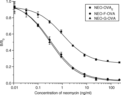
Table II. B0 and IC50 values of ciELISA under optimum concentration of antiserum and coating antigens.
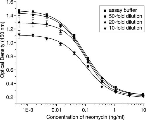
Figure 4. Influence of matrix in milk on the neomycin ELISA standard curve. The dilution times of milk in assay buffer (PBS, pH 7.0) were as follows: control (▪), 50-fold (•), 20-fold (▴), and 10-fold (▾). Each point represents the average of three well replicates, and error bars represent standard deviations.