Figures & data
Table 1. Maize samples analysed in the study.
Table 2. Instrumentation and experimental setup in the participating laboratories.
Table 3. Analysis of normality of data distribution by the Shapiro–Wilk test by sample, laboratory and protocol.
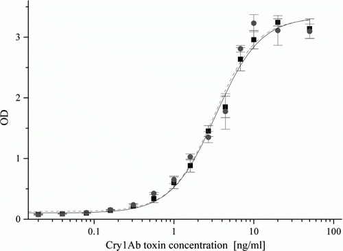
Figure 1. Analytical standard curves obtained in Lab 1 (▪, solid line), Lab 2 (•, dashed line), Lab 3 (![]()
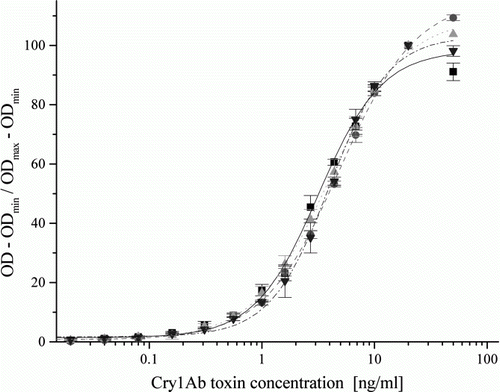
Figure 2. Analytical standard curves obtained in Lab 4 in buffer (▪, solid line) and in isogenic leaf extract (•, dashed line) in the standardised JP. Spiked concentrations of the Cry1Ab toxin standard were 0.01, 0.02, 0.04, 0.08, 0.16, 0.31, 0.56, 1, 1.6, 2.7, 4.4, 6.8, 10, 20 and 50 ng/ml.