Figures & data
Table 1. The affection of different test volume to the test results.
Figure 1. Optimisation of reaction time.
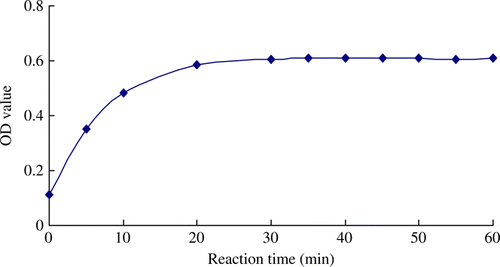
Figure 2. Optimisation of PEG's content. The concentration of IgG standard at 1.0 mg/mL and the concentration of Rabbit anti-bovine IgG at 0.1 mg/mL were selected as working concentration in the optimisation.
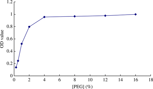
Figure 3. Typical standard curves for IgG immunoassay. Data represent the means of four determinations.
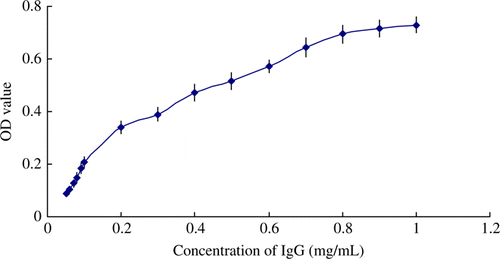
Table 2. OD value of each determination.
Table 3. OD value of each determination.
Table 4. Determinations and recoveries of IgG from spiked milk samples.
Table 5. Determination of IgG's content in liquid bovine colostrum samples.
Table 6. Determination of IgG's content in bovine colostrum powder samples.
Table 7. Repeatability results of the immunoassay.
Table 8. Reproducibility results of the immunoassay.
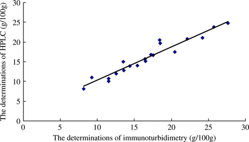
Figure 4. Correlation between IgG concentrations measured by immunoturbidimetry method and by HPLC in bovine colostrum powder samples. y = 0.843x + 1.893, r 2=0.930, n = 20.