866
Views
20
CrossRef citations to date
0
Altmetric
Original Articles

Soybean allergen glycinin induced the destruction of the mechanical barrier function in IPEC-J2

, , , , &
Pages 601-609 | Received 18 Sep 2014, Accepted 26 Nov 2014, Published online: 19 Jan 2015

Figures & data

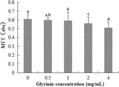
Figure 1. Metabolic activity of IPEC-J2 cells after 24 h incubation of glycinin measured by MTT assay. Each bar represents 3 independent experiments performed in triplicates ± SD. a, b, c, d each represent two groups for different concentrations which are statistically different from each other.
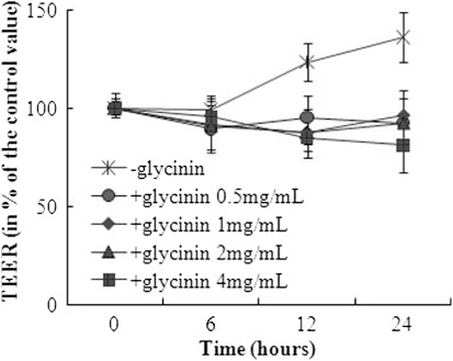
Figure 2. TEER of IPEC-J2 cells after 0, 6, 12 and 24 h incubation of glycinin. Each bar represents four independent experiments performed in triplicates ± SD.
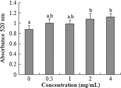
Figure 3. AP activity of IPEC-J2 cells after 72 h incubation of glycinin. Each bar represents four independent experiments performed in triplicates ± SD. a, b each represent two groups for different concentrations which are statistically different from each other.
Figure 4. TJ proteins immunofluorescence of IPEC-J2 cells after 24 h incubation of glycinin. Representative images of immunofluorescence staining (magnification 200×) are shown.
Figure 5. Western blot of TJ proteins of IPEC-J2 cells after 24 h incubation of glycinin. The β-actin is housekeeping protein. Representative Western blots from four independent experiments are shown.

Reprints and Corporate Permissions

Please note: Selecting permissions does not provide access to the full text of the article, please see our help page How do I view content?

To request a reprint or corporate permissions for this article, please click on the relevant link below:

Academic Permissions

Please note: Selecting permissions does not provide access to the full text of the article, please see our help page How do I view content?

Obtain permissions instantly via Rightslink by clicking on the button below:

If you are unable to obtain permissions via Rightslink, please complete and submit this Permissions form. For more information, please visit our Permissions help page.