Figures & data
Figure 1. Comparison of UV spectra of PSL, PSL-21-HS, BSA and PSL-21-HS-BSA.
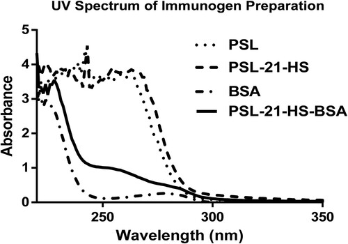
Figure 2. Composite dose–response curve of homologous ELISA of prednisolone using PSL-21-HS-BSA-antibody with PSL-21-HS-HRP-enzyme conjugate. Each value is a mean ± SD of eight assays (In duplicate). The coefficient of variation at each concentration is shown in parentheses.
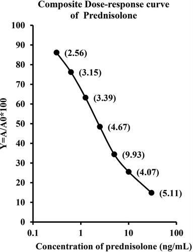
Table 1. Slope (m), Intercept (c), Sensitivity, Affinity, ED50 and R² of prednisolone assay, using PSL-21-HS-BSA-antibody with PSL-21-HS-HRP-enzyme conjugate.
Table 2. Cross-reactivity of steroid compounds with Prednisolone in homologous and bridge heterologous assays of Prednisolone using PSL-21-HS-BSA-antibody with PSL-21-HS-HRP-enzyme conjugates by ELISA.
Table 3. Recovery of prednisolone from exogenously spiked serum pools.
Table 4. Inter- and intra-assay CV % for the measurement of prednisolone in serum pool.
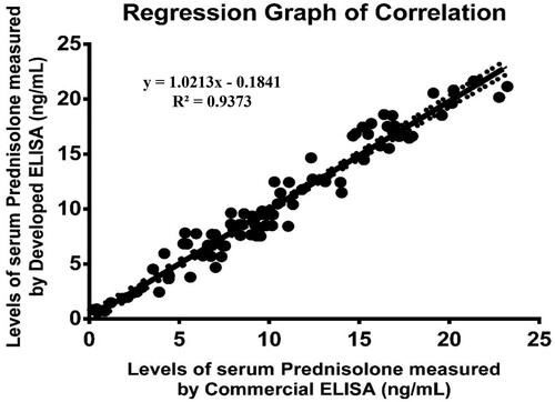
Figure 3. Regression graph of correlation between the serums prednisolone concentrations as estimated by the developed ELISA and an established ELISA kit (plotted by Prism 6.0 software).