Figures & data
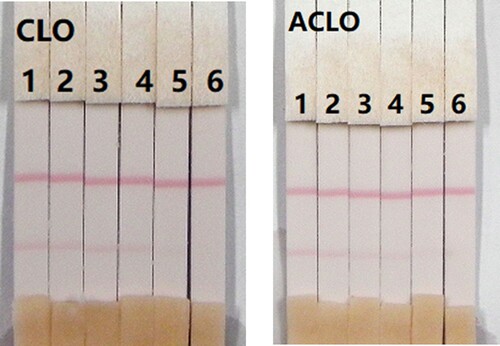
Figure 1. Chemical structure of clonidine (A) and apraclonidine (B).
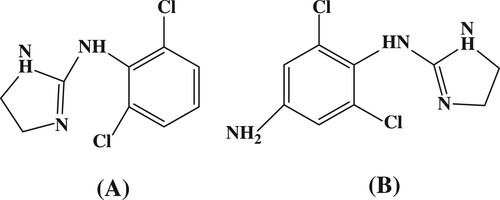
Figure 2. The UV spectra characterization for clonidine (CLO), clonidine was conjugated to OVA by the diazobenzidine method (CLO-DIA-OVA), and OVA.
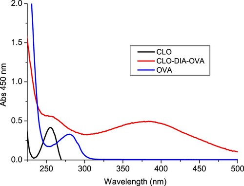
Figure 3. The UV spectra characterization for clonidine (CLO), clonidine was conjugated to KLH by the diazobenzidine method (CLO-DIA-KLH), and KLH.
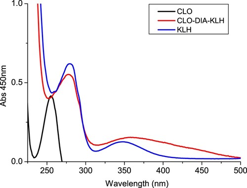
Figure 4. The illustration of strip test results. (A) The sample is negative; (B) if only the C line appears indicates a positive result; (C) invalid result if the control and test line does not appears. C = control line. T = test line.
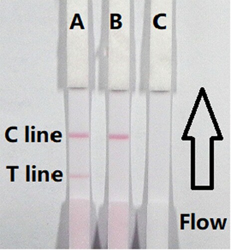
Figure 5. Result of optimization by using 13 kinds of surfactants. 1 = suspension buffer, 2 = PVP, 3 = PEG, 4 = BSA, 5 = Casein, 6 = Sucrose, 7 = Trehalose, 8 = Sorbitol, 9 = Mannitol, 10 = Tween-20, 11 = Brij-35, 12 = Triton X-100, and 13 = On-870. a = negative (0 ng/mL). b = positive (25 ng/mL).
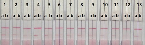
Figure 6. Result of optimization by using two kinds of surfactant and two kinds of coating concentration. A = 0.5 mg/mL of coating concentration; B = 1 mg/mL of coating concentration. a = 0 ng/mL; b = 5 ng/mL.
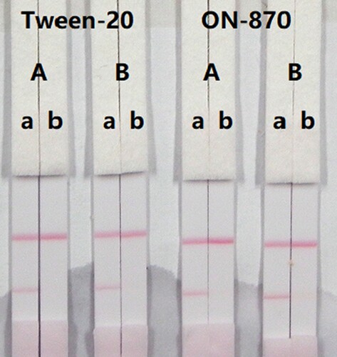
Figure 7. Colloidal gold immunochromatographic strip for CLO in 0.01 M PBS (pH 7.4). CLO concentration: 1 = 0 ng/mL; 2 = 0.1 ng/mL; 3 = 0.25 ng/mL; 4 = 0.5 ng/mL; 5 = 1 ng/mL; and 6 = 2.5 ng/mL.
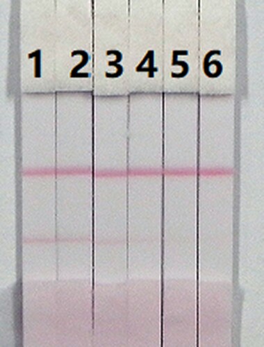
Figure 8. Colloidal gold immunochromatographic strip for ACLO in 0.01 M PBS (pH 7.4). ACLO concentration: 1 = 0 ng/mL; 2 = 0.1 ng/mL; 3 = 0.25 ng/mL; 4 = 0.5 ng/mL; 5 = 1 ng/mL; and 6 = 2.5 ng/mL.
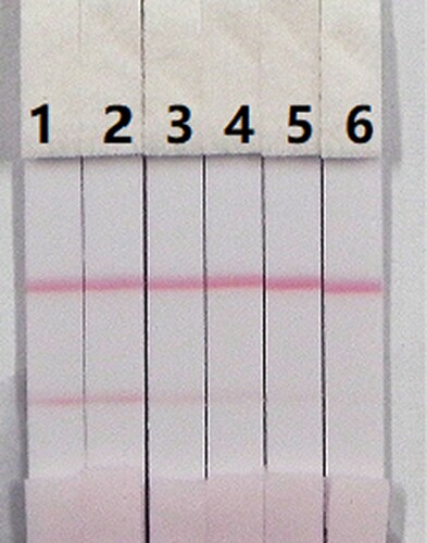
Figure 9. Optimization of the immunochromatographic strip in pig urine. Concentration of coating antigen (A) 0.5 mg/mL and (B) 1 mg/mL. The dosage of the mAb that add in GNP: (a) 8 µg/L and (b) 10 µg/L. The standard concentration: (1) 0 ng/mL and (2) 5 ng/mL.
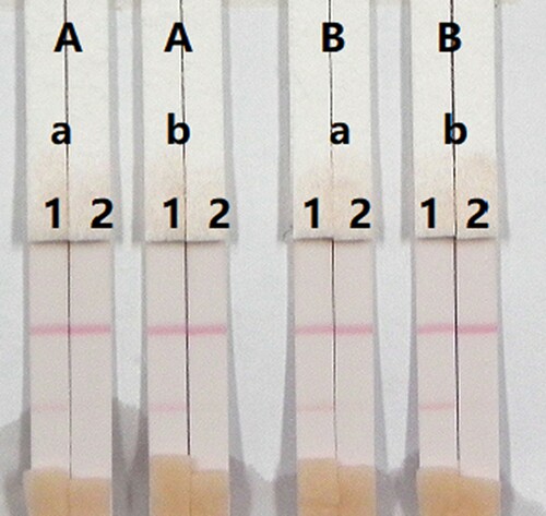
Figure 10. Result of CLO and ACLO spiked in pig urine. CLO and ACLO standard concentration: 1= 0 ng/mL; 2 = 0.1 ng/mL; 3 = 0.25 ng/mL; 4= 0.5 ng/mL; 5 = 1 ng/mL; and 6= 2.5 ng/mL.