Figures & data
Figure 1. VEGF levels are upregulated in cancer patients. VEGF expression levels in tissue samples obtained from patients with cancer and healthy individuals retrieved from the Oncomine dataset. Data represent the mean ± S.E.M.
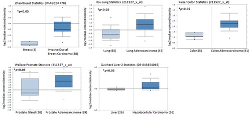
Figure 2. VEGF promotes EPC angiogenesis in vivo. (A) Five-day-old fertilized chick embryos were treated with VEGF. After 3 days, the CAMs were examined by microscopy and photographed. (B) Matrigel plugs were treated with VEGF and subcutaneously injected into the flanks of nude male mice. After 7 days, the plugs were photographed and immunostained with CD31, CD34, and CD133 antibodies. Data represent the mean ± S.E.M. *, p < 0.05 compared with the control group.
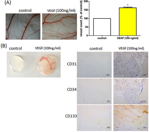
Figure 3. Soya-cerebroside does not induce cell death in EPCs. EPCs were treated with varying concentrations of soya-cerebroside (1–10 μM) for 24 or 48 h and cell viability was determined using the MTT assay. Data represent the mean ± S.E.M.
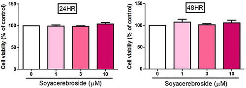
Figure 4. Soya-cerebroside inhibits VEGF-facilitated EPC migration. EPCs were incubated with VEGF and soya-cerebroside (1–10 μM) for 24 h. Cell migration was examined by the Transwell assay. Data represent the mean ± S.E.M. *, p < 0.05 compared with the control group; #, p < 0.05 compared with the VEGF-treated group.
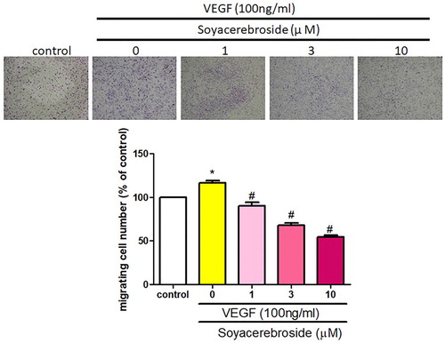
Figure 5. Soya-cerebroside inhibits VEGF-facilitated EPC tube formation. EPCs were incubated with VEGF and soya-cerebroside (1–10 μM) for 24 h. Capillary-like structure formation was examined using the tube formation assay. Data represent the mean ± S.E.M. *, p < 0.05 compared with the control group; #, p < 0.05 compared with the VEGF-treated group.
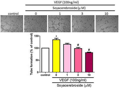
Figure 6. Soya-cerebroside inhibits VEGF-facilitated c-Src and FAK phosphorylation. EPCs were incubated with VEGF and soya-cerebroside (1–10 μM) for 24 h. c-Src, FAK, PI3 K, Akt, Raf and MEK phosphorylation was examined by Western blot analysis.
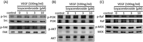
Figure 7. Soya-cerebroside inhibits VEGF-facilitated Runx2 activation. EPCs were incubated with VEGF and soya-cerebroside (1–10 μM) for 24 h. Runx2 and c-Jun expression was examined by Western blot analysis.
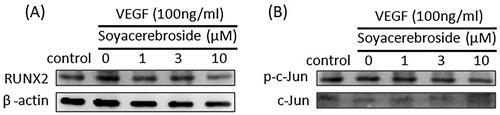
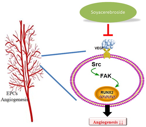
Figure 8. Schema describing the effects of soya-cerebroside in VEGF-promoted EPC angiogenesis. Soya-cerebroside inhibits VEGF-facilitated EPC angiogenesis by inhibiting the c-Src, FAK and Runx2 signalling pathways.