Figures & data
Figure 1. Resultant sequence of GRA1 and GRA7. Antigenic epitopes of GRA1 and GRA7 antigenic epitopes for Toxoplasma gondii were analysed and linked by GG and GS codons. The rare codons of the sequences were replaced with E.coli-preferred codons.
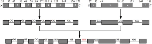
Figure 2. Schematic illustration of GICA strip. The serum is loaded onto the sample pad and the gold–rSPG conjugate is added onto the conjugate pad.rGRA is immobilized as the test line in the NC membrane. The rSPG is used as the control line. Following the application of a serum sample containing specific anti-Toxoplasma gondii IgG and non-specific IgG onto the NC membrane, the conjugated anti-Toxoplasma gondii IgG complex is captured by rGRA on the test line (T), resulting in a red band. The conjugated anti-Toxoplasma gondii IgG and nonspecific IgG are captured by the rSPG on the control line (C), resulting in another red band.
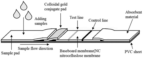
Figure 3. The construction of pET-32a(+)-GRA recombinant plasmid. The sequences of GRA were obtained, TAA were inserted at its 3′ end and BamH1 and EcoR1 restriction sites were inserted at its 5′ and 3′ ends, respectively. The total length of the sequence was 663 bp.
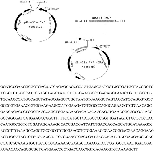
Figure 4. rGRA expression, purification and identification. Expression, purification and identification of rGRA. A: PCR identification of bacteria solution. M: DNA marker. 1: Amplified GRA gene fragment. B: Double-enzyme digestion identification M: DNA marker. 1: Recombinant plasmid digested with restriction enzymes. C: Purification of rGRA. M: Protein marker, 1: Supernatant of the expression product; 2: Precipitate of the expression product; 3: Purified rGRA. D: Western-blot analysis of rGRA. M: Protein marker 1: Purified rGRA recognized with Toxoplasma-positive serum.
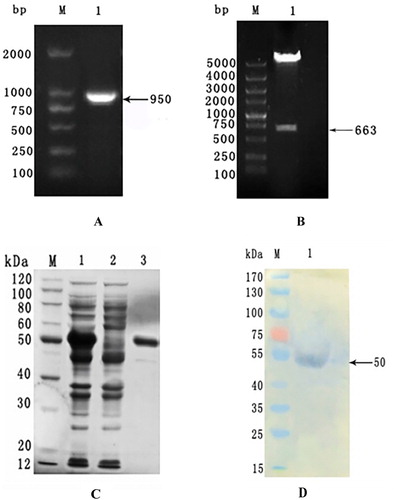
Table 1. Results of ELISA assay on the detection threshold of rGRA.
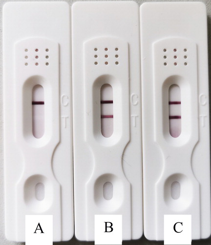
Figure 5 Mouse serum detection using GICA strips. A: Negative result; B: Weakly positive result; C: positive result.