Figures & data
Figure 1. IL-1β, IL-6 and IL-8 levels are upregulated in RA patients. Levels of IL-1β, IL-6 and IL-8 expression in tissue samples retrieved from the GEO dataset, including patients with RA and healthy individuals.
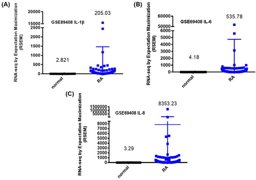
Figure 2. IL-1β, IL-6 and IL-8 levels are upregulated in CIA mouse tissue. Histologic sections of ankle joints were immunostained with IL-1β, IL-6 and IL-8 in healthy control mice and CIA mice.
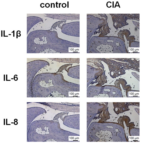
Figure 3. Soya-cerebroside inhibits IL-1β, IL-6 and IL-8 expression in RASFs. (A) RASFs were incubated with varying concentrations of soya-cerebroside (1–10 μM) for 24 h and cell viability was determined using the MTT assay. (B-E) RASFs were treated with soya-cerebroside (1–10 μM) for 24 h, and IL-1β, IL-6 and IL-8 expression were examined by qPCR and Western blot analysis. Data represent the mean ± S.D. *p < 0.05 compared with the control group.
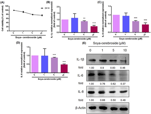
Figure 4. Soya-cerebroside inhibits ERK phosphorylation, but not p38 or JNK phosphorylation. RASFs were incubated with soya-cerebroside (1–10 μM) for 24 h. ERK, p38 and JNK phosphorylation was examined by Western blot analysis.
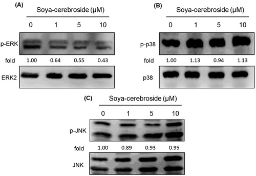
Figure 5. Soya-cerebroside does not inhibit PI3K or Akt phosphorylation. RASFs were incubated with soya-cerebroside (1–10 μM) for 24 h. PI3K and Akt phosphorylation was examined by Western blot analysis.
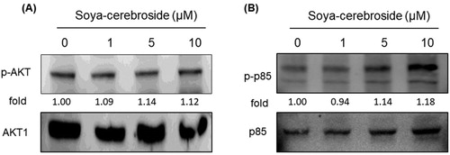
Figure 6. Soya-cerebroside inhibits activation of NF-κB and AP-1. RASFs were incubated with soya-cerebroside (1–10 μM) for 24 h. p65 and c-Jun phosphorylation was examined by Western blot analysis.
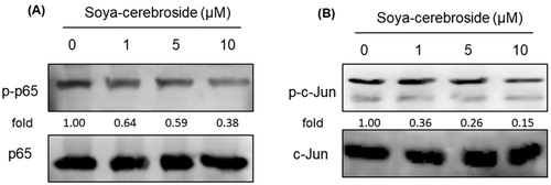
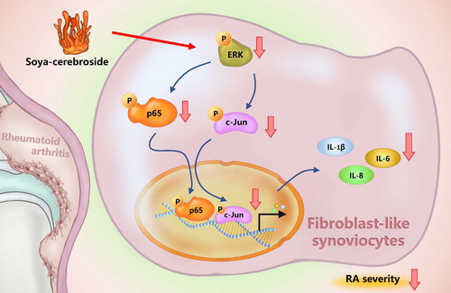
Figure 7. Schema illustrating the effects of soya-cerebroside in IL-1β, IL-6 and IL-8 expression. Soya-cerebroside inhibits IL-1β, IL-6 and IL-8 expression during RA progression by inhibiting the ERK, NF-κB and AP-1 signalling pathways.