Figures & data
Table 1. The sequence of oligonucleotide primers used for RT-PCR.
Figure 1. Effect of APF on macrophage activation in RAW264.7 cells. RAW264.7 cells were treated with APF for 24 h. (A) NO level, (B) cell viability and (C) mRNA level were measured by the Griess assay, MTT assay and RT-PCR, respectively. *p < 0.05 compared to the cells without the treatment.
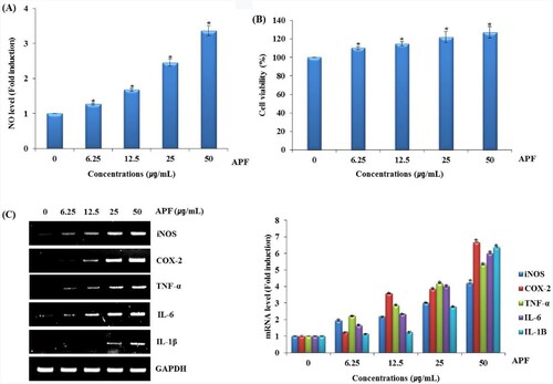
Figure 2. Effect of TLR2/TLR4 on APF-mediated production of immunomodulators in RAW264.7 cells. RAW264.7 cells were pretreated with C29 (TLR2 inhibitor, 10 μM) or TAK-242 (TLR4 inhibitor, 10 μM) for 2 h and co-treated with APF (50 μg/ml) for 24 h. (A) NO level (C29), (B) NO level (TAK-242), and (C) mRNA level (TAK-242) were measured by Griess assay (A and B) and RT-PCR (C), respectively. *p < 0.05 compared to the cells without the treatment.
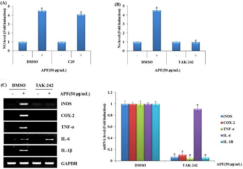
Figure 3. Effect of MAPK signalling pathway on APF-mediated production of immunomodulators in RAW264.7 cells. RAW264.7 cells were pretreated with (A) PD98059 (ERK1/2 inhibitor, 40 μM), (B) SB203580 (p38 inhibitor, 40 μM) or (C) SP600125 (JNK inhibitor, 40 μM) for 2 h and then co-treated with APF (50 μg/ml) for 24 h. NO level was measured by the Griess assay. (D) RAW264.7 cells were pretreated with SP600125 (JNK inhibitor, 40 μM) for 2 h and then co-treated with APF (50 μg/ml) for 24 h. mRNA level was measured by the RT-PCR. (E) RAW264.7 cells were treated with APF (50 μg/ml) for the indicated times. Protein levels were measured by Western blot analysis. (F) RAW264.7 cells were pretreated with TAK-242 (TLR4 inhibitor, 10 μM) for 2 h and co-treated with APF (50 μg/ml) for 1 h. Protein levels were measured by the Western blot analysis. *p < 0.05 compared to the cells without the treatment.
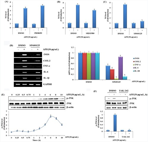
Figure 4. Effect of NF-κB signalling pathway on APF-mediated production of immunomodulators in RAW264.7 cells. (A and B) RAW264.7 cells were pretreated with BAY 11–7082 (NF-κB inhibitor, 20 μM) for 2 h and then co-treated with APF (50 μg/ml) for 24 h. NO level was measured by the Griess assay and RT-PCR, respectively. (C) RAW264.7 cells were treated with APF (50 μg/ml) for the indicated times. Protein levels were measured by Western blot analysis. (D) RAW264.7 cells were pretreated with TAK-242 (TLR4 inhibitor, 10 μM) for 2 h and co-treated with APF (50 μg/ml) for 1 h. Protein levels were measured by the Western blot analysis. *p < 0.05 compared to the cells without the treatment.
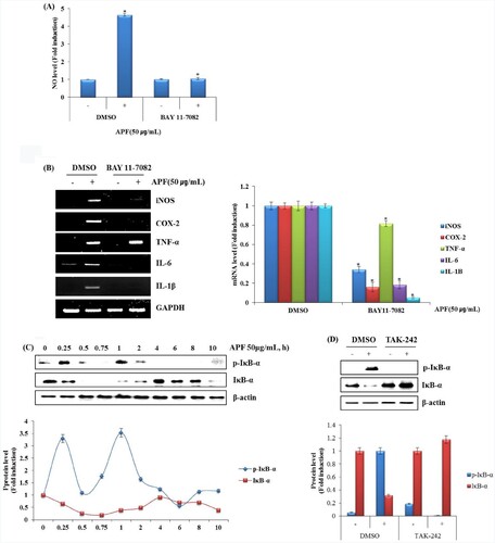
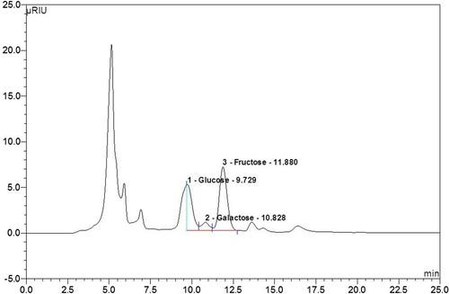
Figure 5. The sugars contained in the polysaccharides of APF were analyzed using high-performance liquid chromatography (HPLC).