Figures & data
Figure 1. Curcumin inhibits the proliferation, invasion, and migration of hepatocellular carcinoma cells. (A, B) Cell growth rates of HepG2 and SK-Hep-1 cells treated with curcumin (10, 20, 40, and 60 µM) and 5FU (10 µM) were measured by CCK-8 assays at 24, 48, and 72 h. (C, D) Semi-quantitative analysis of the in vitro migration ability of HepG2 and SK-Hep-1 cells treated with curcumin (20, 40, and 60 µM) and 5FU (10 µM) at 24 h was performed by a scratch healing assay (×100). (E, F) Transwell assays were used to assess the invasive ability of HepG2 and SK-Hep-1 cells treated with curcumin (20, 40, and 60 µM) and 5FU (10 µM) at 24 h. The number of invaded cells was analyzed (×200). (G)The expression of invasion and metastasis-related proteins MMP2 and MMP9 was quantitated by western blotting. **P < 0.01.
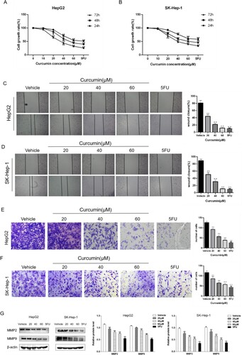
Figure 2. Curcumin inhibits Wnt/β-catenin signalling in hepatocellular carcinoma cells. (A) Western blotting was used to detect the expression of Wnt/β-catenin pathway-related proteins in HepG2 cells and SK-Hep-1 treated with curcumin (20, 40, and 60 µM) and 5FU (10 µM) at 24 h. (B) Western blot analysis of β-catenin TCF4, c-Myc, MMP2, and MMP9 in HepG2 cells and SK-Hep-1 cells after addition of PKF-118-310. Each group compared with the vehicle group, **P < 0.01, PKF-118-310 group compared with curcumin + PKF-118-310 group, ##P < 0.01.
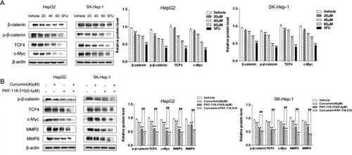
Figure 3. Curcumin regulates Bclaf1 to affect the invasion and metastasis of HCC cells. (A) HepG2 and SK-Hep-1 cells were treated with curcumin (20, 40, and 60 µM) and 5-FU (10 µM) for 24 h and then Bclaf1 protein expression was detected by western blotting. (B) Western blotting was used to assess the effects of curcumin (40 µM) on p-β-catenin, TCF4, c-Myc, MMP2, and MMP9 protein expression in HepG2 cells after Bclaf1 knockout. (C, D) Semi-quantitative analysis of the effect of curcumin (40 µM) on HepG2 and SK-Hep-1 cell migration after Bclaf1 knockout (×100) and the healing rate of scratch wounds. (E, F) Transwell assays were used to determine the effect of curcumin (40 µM) on HepG2 cell invasion after Bclaf1 knockout in vitro (×200). The number of invaded cells was analyzed. Each group compared with the blank control group, **P < 0.01, curcumin group compared with the sgRNA+ curcumin group, ##P < 0.01.
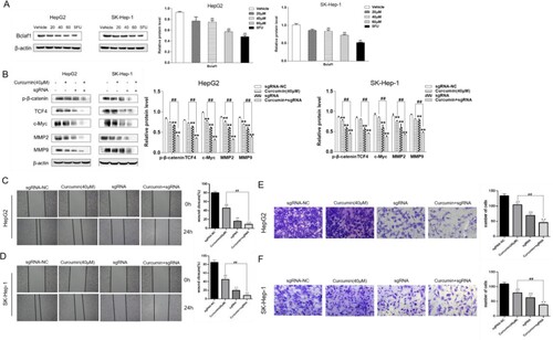
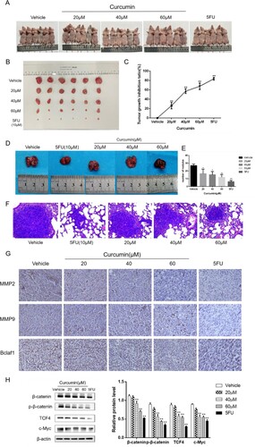
Figure 4. Curcumin has anti-tumour and -metastasis effects on subcutaneously transplanted tumours in nude mice. (A, B) Mice were sacrificed by cervical dislocation and subcutaneous tumour tissues were observed and photographed. (C). Inhibitory effect of curcumin on transplanted tumours in nude mice of each experimental group. (D, E) After sacrificing mice, lungs were collected, photographed, and fixed, and the formation of lung tumour nodules was compared between the experimental groups. (F) Lung tissue sections were observed after HE staining and photographed (×100). (G) Effect of curcumin on MMP2, MMP9, and Bclaf1 expression in HepG2 xenograft tissues in nude mice was detected by immunohistochemistry (×400). (H) Western blot analysis of curcumin effects on Wnt/β-catenin pathway protein expression in transplanted tumour tissue in nude mice. Each group compared with the vehicle group, **P < 0.01.