
Figures & data
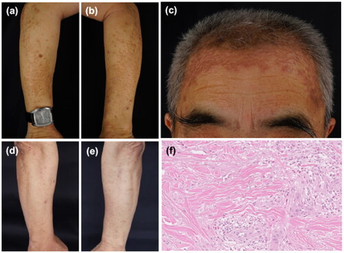
Figure 1. (a–c) Clinical presentation before treatment. Yellow-red papules and plaques on the forearms and on the forehead. (d,e) A clinical picture on the forearms after 2 weeks of treatment. (f) Histological analysis from the left forearm with hematoxylin and eosin staining (×200) showed palisade-like granulomas in the dermis, accompanied by eosinophil infiltration.
Data availability statement
The data that support the findings are available from the corresponding author, Aiping Wang, upon reasonable request.