
Figures & data
Figure 1. Erythematous plaques covered by multiple pustules in a 33-year-old female (). Histopathological picture of an incisional biopsy from a plaque on the back. Section of the skin characterized by epidermal hyperplasia, orto-parakeratosis, mixed inflammatory infiltrate in the dermis (), and micro pustules (). (c) Hematoxylin and Eosin stain, ×10 magnification; (d) Hematoxylin and Eosin stain, ×20 magnification
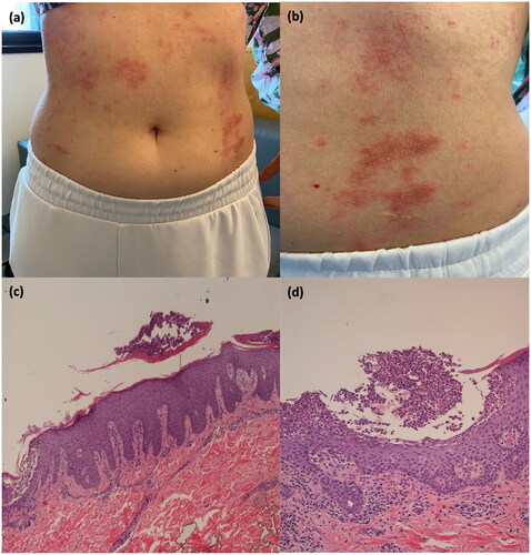
Table 1. Evaluation of causality relation according to Naranjo algorithm.
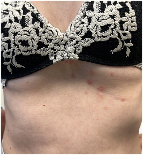
Figure 2. Almost complete remission of pustular psoriasis after 4 weeks of follow-up.
Data availability statement
Data available on request from the authors.