Figures & data
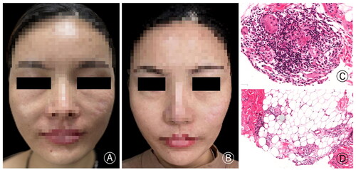
Figure 1. (A) Nodules and swelling developed in the bilateral infraorbital region, particularly on the right side. (B) The symptoms were significantly relieved after treatment. (C) Foreign bodies were observed dispersed among inflammatory cells, epithelioid cells, and macrophages. Additionally, multinucleated foreign body giant cells were also observed (H&E × 200).
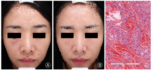
Figure 2. (A) The patient had erythema and swelling in the central area of the face, especially in the bilateral suborbital and chin regions. (B) The symptoms significantly improved after approximately one month of treatment. (C) MRI (T2W) coronal image revealed the presence of enhancing signal and space-occupying changes with indistinct margins.
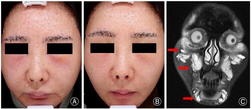
Figure 3. (A) The patient had swelling and nodules at the bilateral nasolabial fold. (B) The symptoms significantly improved after treatment. (C) The histopathology showed foreign body granuloma composed of more lymphocytes, macrophages and injected foreign bodies, and there was also inflammatory cell infiltrating around the adipose tissue (H&E × 200).