Figures & data
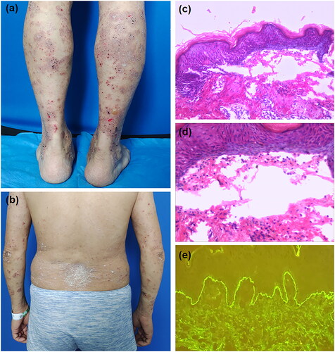
Figure 1. (a) Scattered thick-walls vesicles based on erythematous on the lower limbs, with some small superficial erosions and crusts. (b) a few scattered red plaques covered with silvery white scales are seen on the back and upper limbs. (c) Histopathology examination shows subepidermal blisters. (d) Small number of eosinophils were seen within the blisters. (e) Direct immunofluorescence studies reveal linear deposition of IgG at the basement membrane zone.