5,198
Views
17
CrossRef citations to date
0
Altmetric
Reviews

Ionizing radiation-induced risks to the central nervous system and countermeasures in cellular and rodent models

, , ORCID Icon & ORCID Icon
Pages S132-S150 | Received 05 Jun 2019, Accepted 11 Feb 2020, Published online: 20 Oct 2020

Figures & data

Figure 1. Summary representation of the discussed countermeasures. Numbers refer to the following studies: 1. (Nair and Nair Citation2013b), 2. (Mohammad et al. Citation2014), 3. (Wang et al. Citation2018), 4. (Guelman et al. Citation2003), 5. (Lamproglou et al. Citation2003), 6. (Guelman et al. Citation2005), 7. (Erol et al. Citation2004), 8. (Limoli et al. Citation2007), 9. (Manda et al. Citation2008), 10. (Leu et al. Citation2017), 11. (Villasana et al. Citation2013), 12. (Allen et al. Citation2014), 13. (Lu et al. Citation2018), 14. (Parihar et al. Citation2015), 15. (Chmielewski et al. Citation2016), 16. (Weitzel et al. Citation2015), 17. (Raber et al. Citation2017), 18.(Ündeğer et al. Citation2004), 19. (El-Missiry et al. Citation2018), 20. (Manda et al. Citation2007), 21. (Facchino et al. Citation2010), 22. (Yang et al. Citation2009), 23. (Yang et al. Citation2011), 24. (Jiang, Perez-Torres, et al. Citation2014), 25. (Pena et al. Citation2000), 26. (El-Missiry et al. Citation2018), 27. (Andratschke et al. Citation2004), 28. (Nieder et al. Citation2006), 29. (Nieder, Andratschke, et al. Citation2005), 30. (Nieder, Price, et al. Citation2005), 31. (Jiang, Perez-Torres, et al. Citation2014), 32. (Gonzalez et al. Citation2007), 33. (Jiang, Engelbach, et al. Citation2014), 34. (Belarbi et al. Citation2013), 35. (Yang et al. Citation2018), 36. (Erbayraktar et al. Citation2006), 37. (Ansari et al. Citation2007), 38. (Yuan et al. Citation2003), 39. (Zhao et al. Citation2007), 40. (Schnegg et al. Citation2012), 41. (Greene-Schloesser et al. Citation2014), 42. (Desmarais et al. Citation2015), 43. (Kim et al. Citation2004), 44. (Lee et al. Citation2012), 45. (Tikka et al. Citation2001), 46. (Feng et al. Citation2016), 47. (Feng et al. Citation2018), 48. (Baulch et al. Citation2016), 49. (Smith et al. Citation2020), 50. (Liao et al. Citation2017), 51. (Wang et al. Citation2016), 52. (Piao et al. Citation2015), 53. (Zhou et al. Citation2015), 54. (Bala et al. Citation2017), 55. (Oh et al. Citation2013), 56. (Sun et al. Citation2013), 57. (Prager et al. Citation2016).

Figure 1. Summary representation of the discussed countermeasures. Numbers refer to the following studies: 1. (Nair and Nair Citation2013b), 2. (Mohammad et al. Citation2014), 3. (Wang et al. Citation2018), 4. (Guelman et al. Citation2003), 5. (Lamproglou et al. Citation2003), 6. (Guelman et al. Citation2005), 7. (Erol et al. Citation2004), 8. (Limoli et al. Citation2007), 9. (Manda et al. Citation2008), 10. (Leu et al. Citation2017), 11. (Villasana et al. Citation2013), 12. (Allen et al. Citation2014), 13. (Lu et al. Citation2018), 14. (Parihar et al. Citation2015), 15. (Chmielewski et al. Citation2016), 16. (Weitzel et al. Citation2015), 17. (Raber et al. Citation2017), 18.(Ündeğer et al. Citation2004), 19. (El-Missiry et al. Citation2018), 20. (Manda et al. Citation2007), 21. (Facchino et al. Citation2010), 22. (Yang et al. Citation2009), 23. (Yang et al. Citation2011), 24. (Jiang, Perez-Torres, et al. Citation2014), 25. (Pena et al. Citation2000), 26. (El-Missiry et al. Citation2018), 27. (Andratschke et al. Citation2004), 28. (Nieder et al. Citation2006), 29. (Nieder, Andratschke, et al. Citation2005), 30. (Nieder, Price, et al. Citation2005), 31. (Jiang, Perez-Torres, et al. Citation2014), 32. (Gonzalez et al. Citation2007), 33. (Jiang, Engelbach, et al. Citation2014), 34. (Belarbi et al. Citation2013), 35. (Yang et al. Citation2018), 36. (Erbayraktar et al. Citation2006), 37. (Ansari et al. Citation2007), 38. (Yuan et al. Citation2003), 39. (Zhao et al. Citation2007), 40. (Schnegg et al. Citation2012), 41. (Greene-Schloesser et al. Citation2014), 42. (Desmarais et al. Citation2015), 43. (Kim et al. Citation2004), 44. (Lee et al. Citation2012), 45. (Tikka et al. Citation2001), 46. (Feng et al. Citation2016), 47. (Feng et al. Citation2018), 48. (Baulch et al. Citation2016), 49. (Smith et al. Citation2020), 50. (Liao et al. Citation2017), 51. (Wang et al. Citation2016), 52. (Piao et al. Citation2015), 53. (Zhou et al. Citation2015), 54. (Bala et al. Citation2017), 55. (Oh et al. Citation2013), 56. (Sun et al. Citation2013), 57. (Prager et al. Citation2016).

Figure 2. Representation of radiation-induced responses of the CNS. [1–4] = (Belka et al. Citation2001; Satyamitra et al. Citation2007; Lowe et al. Citation2009; Baluchamy et al. Citation2010; Beckhauser et al. Citation2016), [5–16] = (Fournier and Taucher-Scholz Citation2004; Limoli et al. Citation2004; Kim et al. Citation2006; Al-Jahdari et al. Citation2008; Eriksson and Stigbrand Citation2010; Chakraborti et al. Citation2012; Parihar and Limoli Citation2013; Shirai et al. Citation2013; Kempf et al. Citation2014; Parihar et al. Citation2015), [17–24] = (Clatworthy et al. Citation1999; Sannita et al. Citation2007; Machida et al. Citation2010; Sanchez et al. Citation2010; Marty et al. Citation2014; Rudobeck et al. Citation2014; Sokolova et al. Citation2015), [25–46] = (Tofilon and Fike Citation2000; Vazquez and Kirk Citation2000; van Vulpen et al. Citation2002; Maier Citation2003; Mizumatsu et al. Citation2003; Lyubimova and Hopewell Citation2004; Raber et al. Citation2004; Rola et al. Citation2004; Casadesus et al. Citation2005; Rola et al. Citation2005; Hwang et al. Citation2006; Fike et al. Citation2009; Huang et al. Citation2010; Moravan et al. Citation2011; Kadir et al. Citation2012; Monje and Dietrich Citation2012; York et al. Citation2012; Rivera et al. Citation2013; Greene-Schloesser et al. Citation2014; Morganti et al. Citation2014; Hur and Yoon Citation2017), [47–60] = (Brouwers and Poplack Citation1990; Hall et al. Citation2004; Butler and Haser Citation2006; Rosi et al. Citation2008; Zeltzer et al. Citation2009; Liu et al. Citation2010; Britten et al. Citation2012; Greene-Schloesser and Robbins Citation2012; Armstrong et al. Citation2013; Greene-Schloesser, Moore, and Robbins Citation2013; Kumar et al. Citation2013; Britten et al. Citation2014; Makale et al. Citation2017; Acharya et al. Citation2019).

Figure 2. Representation of radiation-induced responses of the CNS. [1–4] = (Belka et al. Citation2001; Satyamitra et al. Citation2007; Lowe et al. Citation2009; Baluchamy et al. Citation2010; Beckhauser et al. Citation2016), [5–16] = (Fournier and Taucher-Scholz Citation2004; Limoli et al. Citation2004; Kim et al. Citation2006; Al-Jahdari et al. Citation2008; Eriksson and Stigbrand Citation2010; Chakraborti et al. Citation2012; Parihar and Limoli Citation2013; Shirai et al. Citation2013; Kempf et al. Citation2014; Parihar et al. Citation2015), [17–24] = (Clatworthy et al. Citation1999; Sannita et al. Citation2007; Machida et al. Citation2010; Sanchez et al. Citation2010; Marty et al. Citation2014; Rudobeck et al. Citation2014; Sokolova et al. Citation2015), [25–46] = (Tofilon and Fike Citation2000; Vazquez and Kirk Citation2000; van Vulpen et al. Citation2002; Maier Citation2003; Mizumatsu et al. Citation2003; Lyubimova and Hopewell Citation2004; Raber et al. Citation2004; Rola et al. Citation2004; Casadesus et al. Citation2005; Rola et al. Citation2005; Hwang et al. Citation2006; Fike et al. Citation2009; Huang et al. Citation2010; Moravan et al. Citation2011; Kadir et al. Citation2012; Monje and Dietrich Citation2012; York et al. Citation2012; Rivera et al. Citation2013; Greene-Schloesser et al. Citation2014; Morganti et al. Citation2014; Hur and Yoon Citation2017), [47–60] = (Brouwers and Poplack Citation1990; Hall et al. Citation2004; Butler and Haser Citation2006; Rosi et al. Citation2008; Zeltzer et al. Citation2009; Liu et al. Citation2010; Britten et al. Citation2012; Greene-Schloesser and Robbins Citation2012; Armstrong et al. Citation2013; Greene-Schloesser, Moore, and Robbins Citation2013; Kumar et al. Citation2013; Britten et al. Citation2014; Makale et al. Citation2017; Acharya et al. Citation2019).

Table 1. Summary of the discussed countermeasure agents classified into five main approaches (targeting reactive oxygen species, DNA damage, cell survival, inflammation and tissue repair), according to the type of CNS impairment to be treated.
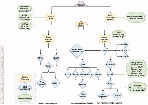
Figure 3. Flowchart of the main discussed processes of radiation-induced targeted and non-targeted DNA damage effects, and associated countermeasures. RIBE: radiation-induced bystander effects, CNPs: anti-histone antibody complexed nanoparticles, R-Cu: resveratrol-copper, ROS: reactive oxygen species, RNS: reactive nitrogen species, SSBs: single-stranded breaks, DSBs: double-stranded breaks, DDR: DNA damage response, BER: base excision repair, HR: homologous recombination, NHEJ: non-homologous end joining. Numbers refer to the following studies: 18. (Ündeğer et al. Citation2004), 19. (El-Missiry et al. Citation2018), 20. (Manda et al. Citation2007), 21. (Facchino et al. Citation2010), 22. (Yang et al. Citation2009), 23. (Yang et al. Citation2011), 24. (Jiang et al. Citation2014) Supplementary Table 2 provides additional information regarding the model, irradiation, administration conditions and main results for each of the discussed countermeasures.

Figure 3. Flowchart of the main discussed processes of radiation-induced targeted and non-targeted DNA damage effects, and associated countermeasures. RIBE: radiation-induced bystander effects, CNPs: anti-histone antibody complexed nanoparticles, R-Cu: resveratrol-copper, ROS: reactive oxygen species, RNS: reactive nitrogen species, SSBs: single-stranded breaks, DSBs: double-stranded breaks, DDR: DNA damage response, BER: base excision repair, HR: homologous recombination, NHEJ: non-homologous end joining. Numbers refer to the following studies: 18. (Ündeğer et al. Citation2004), 19. (El-Missiry et al. Citation2018), 20. (Manda et al. Citation2007), 21. (Facchino et al. Citation2010), 22. (Yang et al. Citation2009), 23. (Yang et al. Citation2011), 24. (Jiang et al. Citation2014) Supplementary Table 2 provides additional information regarding the model, irradiation, administration conditions and main results for each of the discussed countermeasures.

Figure 4. Flowchart of the main discussed processes of radiation-induced cell cycle modifications, and associated countermeasures. AMPK: AMP-activated protein kinase, JNK: Jun N-terminal kinase, BAX: Bcl-2-associated X protein, BAD: Bcl-2-associated death promoter protein, TSC2: tuberous sclerosis complex-2, mTOR: mammalian target of rapamycin, ASM: acid sphingomyelinase, PI3K: phosphatidylinositol-3 kinase, Akt: serine/threonine-specific protein kinase, PKC: Protein kinase C, IGF-1: insulin-like growth factor-1, MAPK: mitogen-activated protein kinase, ERK: extracellular signal-regulated kinase, EGCG: epigallocathechin-3-gallate, FGF: fibroblast growth factor, PDGF: platelet-derived growth factor. Numbers refer to the following studies: 25. (Pena et al. Citation2000), 26. (El-Missiry et al. Citation2018), 27. (Andratschke et al. Citation2004), 28. (Nieder et al. Citation2006), 29. (Nieder, Andratschke, et al. Citation2005), 30. (Nieder, Price, et al. Citation2005) Supplementary Table 3 provides additional information regarding the model, irradiation, administration conditions and main results for each of the discussed countermeasures.

Figure 4. Flowchart of the main discussed processes of radiation-induced cell cycle modifications, and associated countermeasures. AMPK: AMP-activated protein kinase, JNK: Jun N-terminal kinase, BAX: Bcl-2-associated X protein, BAD: Bcl-2-associated death promoter protein, TSC2: tuberous sclerosis complex-2, mTOR: mammalian target of rapamycin, ASM: acid sphingomyelinase, PI3K: phosphatidylinositol-3 kinase, Akt: serine/threonine-specific protein kinase, PKC: Protein kinase C, IGF-1: insulin-like growth factor-1, MAPK: mitogen-activated protein kinase, ERK: extracellular signal-regulated kinase, EGCG: epigallocathechin-3-gallate, FGF: fibroblast growth factor, PDGF: platelet-derived growth factor. Numbers refer to the following studies: 25. (Pena et al. Citation2000), 26. (El-Missiry et al. Citation2018), 27. (Andratschke et al. Citation2004), 28. (Nieder et al. Citation2006), 29. (Nieder, Andratschke, et al. Citation2005), 30. (Nieder, Price, et al. Citation2005) Supplementary Table 3 provides additional information regarding the model, irradiation, administration conditions and main results for each of the discussed countermeasures.

Figure 5. Flowchart of the main discussed processes of radiation-induced inflammatory responses, and associated countermeasures. EPO: erythropoietin, HIF-1α: hypoxia-inducible factor α, VEGF: vascular endothelial growth factor, CXCR4: CXC motif chemokine receptor 4, CCR2: CC motif chemokine receptor 2, ACE: angiotensin-converting enzyme, Ang II: angiotensin II, TNF-α: tumor necrosis factor α, VCAM-1: vascular cell adhesion protein 1, ICAM-1: intercellular adhesion molecule 1, GSK-3: glycogen synthase kinase 3, DAMPs: danger-associated molecular patterns, TLRs: toll-like receptors, NF-κB: nuclear factor NF-κB, AP-1: activator protein 1, PPAR: proliferator-activated receptor, COX-2: cyclooxygenase-2, iNOS: inducible nitric oxide synthase, NLRP3: NLR Family Pyrin Domain Containing 3, IL-1: interleukin-1, IFN-γ: interferon-γ, TGF-β: transforming growth factor β. Numbers refer to the following studies: 31. (Jiang, Perez-Torres, et al. Citation2014), 32. (Gonzalez et al. Citation2007), 33. (Jiang, Engelbach, et al. Citation2014), 34. (Belarbi et al. Citation2013), 35. (Yang et al. Citation2018), 36. (Erbayraktar et al. Citation2006), 37. (Ansari et al. Citation2007), 38. (Yuan et al. Citation2003), 39. (Zhao et al. Citation2007), 40. (Schnegg et al. Citation2012), 41. (Greene-Schloesser et al. Citation2014), 42. (Desmarais et al. Citation2015), 43. (Kim et al. Citation2004), 44. (Lee et al. Citation2012) Supplementary Table 4 provides additional information regarding the model, irradiation, administration conditions and main results for each of the discussed countermeasures.

Figure 5. Flowchart of the main discussed processes of radiation-induced inflammatory responses, and associated countermeasures. EPO: erythropoietin, HIF-1α: hypoxia-inducible factor α, VEGF: vascular endothelial growth factor, CXCR4: CXC motif chemokine receptor 4, CCR2: CC motif chemokine receptor 2, ACE: angiotensin-converting enzyme, Ang II: angiotensin II, TNF-α: tumor necrosis factor α, VCAM-1: vascular cell adhesion protein 1, ICAM-1: intercellular adhesion molecule 1, GSK-3: glycogen synthase kinase 3, DAMPs: danger-associated molecular patterns, TLRs: toll-like receptors, NF-κB: nuclear factor NF-κB, AP-1: activator protein 1, PPAR: proliferator-activated receptor, COX-2: cyclooxygenase-2, iNOS: inducible nitric oxide synthase, NLRP3: NLR Family Pyrin Domain Containing 3, IL-1: interleukin-1, IFN-γ: interferon-γ, TGF-β: transforming growth factor β. Numbers refer to the following studies: 31. (Jiang, Perez-Torres, et al. Citation2014), 32. (Gonzalez et al. Citation2007), 33. (Jiang, Engelbach, et al. Citation2014), 34. (Belarbi et al. Citation2013), 35. (Yang et al. Citation2018), 36. (Erbayraktar et al. Citation2006), 37. (Ansari et al. Citation2007), 38. (Yuan et al. Citation2003), 39. (Zhao et al. Citation2007), 40. (Schnegg et al. Citation2012), 41. (Greene-Schloesser et al. Citation2014), 42. (Desmarais et al. Citation2015), 43. (Kim et al. Citation2004), 44. (Lee et al. Citation2012) Supplementary Table 4 provides additional information regarding the model, irradiation, administration conditions and main results for each of the discussed countermeasures.
Supplemental material

SupplementaryTable5.docx

Download MS Word (25.6 KB)

SupplementaryTable4.docx

Download MS Word (30.3 KB)

SupplementaryTable3.docx

Download MS Word (19.9 KB)

SupplementaryTable2.docx

Download MS Word (21.7 KB)

SupplementaryTable1.docx

Download MS Word (31.8 KB)