Figures & data
Figure 1. Set up used for imaging procedure, with markings for the proximal (30) and distal (70) imaging sites.
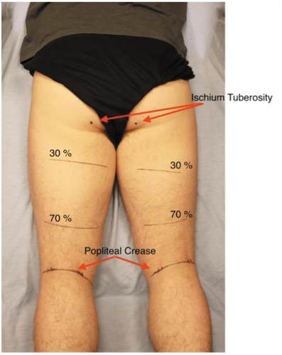
Table 1. Mean, standard deviation (SD) and range for demographic variables.
Figure 2. Mean and standard deviation (SD) values for cross-sectional area (CSA (cm2) of semitendinosus (ST) and the long head of biceps femoris (BF) at proximal (30) and distal (70) sites for the injured (INJ) and sound (SND) limbs of participants with history of anterior cruciate ligament reconstruction (ACLR) and controls (CTRL) with random limb assignment.
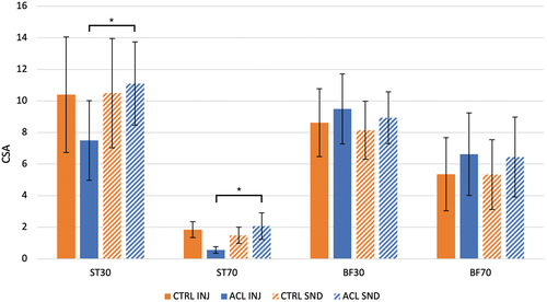
Table 2. Mean and standard deviation (SD) values for cross-sectional area (CSA (cm2) measurements for semitendinosus (ST) and the long head of biceps femoris (BF) for the injured (INJ) and sound (SND) limbs of participants with history of anterior cruciate ligament reconstruction (ACLR) and controls (CTRL) with random limb assignment performed at 30 and 70% of the distance from the ischial tuberosity to the popliteal crease.
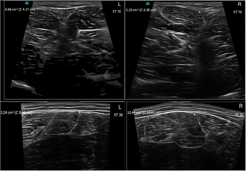
Figure 3. Ultrasound images of a participant from the ACLR group showing the injured (L) and sound (R) limb, with outline tracings of the semitendinosus (ST) for the distal (70; above) and proximal (30; below) sites.