943
Views
42
CrossRef citations to date
0
Altmetric
Original Articles

Systematic reassessment of the red algal genus Phyllymenia (Halymeniaceae, Rhodophyta)

, , , &
Pages 169-178 | Received 19 Jul 2004, Accepted 03 Feb 2005, Published online: 20 Feb 2007

Figures & data

Table 1.  List of species used in rbcL analysis with accession numbers

Figs 1–3.Phyllymenia types and external morphology. . Type sheet of Phyllymenia belangeri (Herb. Bornet & Thuret, PC TA14583). Top left– young specimen of Gigartina polycarpa; bottom left–basal part is Sarcothalia stiriata and distal blade represents P. belangeri (insert); right–female gametophyte of P. belangeri. . Lectotype of Phyllymenia hieroglyphica (LD 22831). . Recently collected specimens from the Western Cape Province, South Africa (GENT TS425) to show habit.
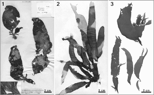
Figs 1–3. Phyllymenia types and external morphology. Fig. 1. Type sheet of Phyllymenia belangeri (Herb. Bornet & Thuret, PC TA14583). Top left– young specimen of Gigartina polycarpa; bottom left–basal part is Sarcothalia stiriata and distal blade represents P. belangeri (insert); right–female gametophyte of P. belangeri. Fig. 2. Lectotype of Phyllymenia hieroglyphica (LD 22831). Fig. 3. Recently collected specimens from the Western Cape Province, South Africa (GENT TS425) to show habit.

Figs 4–10.Phyllymenia vegetative morphology. . Detail of the characteristic corrugated surface with irregular perforations (arrowheads). . Cross-section showing gradual transition between the cortex and medullary layer. . Longitudinal section through a tetrasporic thallus showing predominantly periclinal arrangement of the medullary filaments. . Cross-section through the basal part of the thallus showing a dense medulla with numerous rhizoidal filaments. , . Detail of cortex with anticlinal rows of dichotomously branched filaments. . Inner cortical cells with lateral protuberances establishing secondary pit connections.
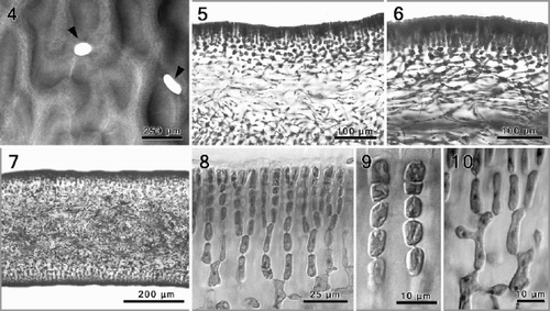
Figs 4–10. Phyllymenia vegetative morphology. Fig. 4. Detail of the characteristic corrugated surface with irregular perforations (arrowheads). Fig. 5. Cross-section showing gradual transition between the cortex and medullary layer. Fig. 6. Longitudinal section through a tetrasporic thallus showing predominantly periclinal arrangement of the medullary filaments. Fig. 7. Cross-section through the basal part of the thallus showing a dense medulla with numerous rhizoidal filaments. Figs 8, 9. Detail of cortex with anticlinal rows of dichotomously branched filaments. Fig. 10. Inner cortical cells with lateral protuberances establishing secondary pit connections.

Figs 11–16.Phyllymenia reproductive morphology. . A maturing cystocarp surrounded by involucral filaments, deeply embedded in the inner cortical layers. . Squashed carpogonial ampulla, with an 8-celled primary filament from which secondary filaments have developed from the first and fifth cell (black arrows), the hypogenous cell (hy) bears a 4-celled filament (white arrow). . A narrowly flask-shaped auxiliary cell ampulla with three, simple secondary filaments. , . Successive stages of gonimoblast development, and formation of involucral filaments; note connecting filaments (cf) attached to the auxiliary cell (ac). . Detail of cruciately divided tetrasporangium and tetrasporangial initials (arrowhead) within modified outer cortical cells.
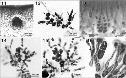
Figs 11–16. Phyllymenia reproductive morphology. Fig. 11. A maturing cystocarp surrounded by involucral filaments, deeply embedded in the inner cortical layers. Fig. 12. Squashed carpogonial ampulla, with an 8-celled primary filament from which secondary filaments have developed from the first and fifth cell (black arrows), the hypogenous cell (hy) bears a 4-celled filament (white arrow). Fig. 13. A narrowly flask-shaped auxiliary cell ampulla with three, simple secondary filaments. Figs 14, 15. Successive stages of gonimoblast development, and formation of involucral filaments; note connecting filaments (cf) attached to the auxiliary cell (ac). Fig. 16. Detail of cruciately divided tetrasporangium and tetrasporangial initials (arrowhead) within modified outer cortical cells.

Fig. 17. Phylogram of one out of two most parsimonious trees (Length = 1163) inferred from rbcL sequence data. Bootstrap percentages of MP (top) and ML (middle) as well as posterior probabilities of BI (bottom) are indicated for each node if higher than 50%.
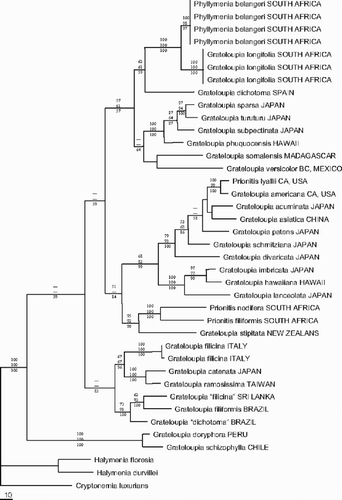
Fig. 17. Phylogram of one out of two most parsimonious trees (Length = 1163) inferred from rbcL sequence data. Bootstrap percentages of MP (top) and ML (middle) as well as posterior probabilities of BI (bottom) are indicated for each node if higher than 50%.

Reprints and Corporate Permissions

Please note: Selecting permissions does not provide access to the full text of the article, please see our help page How do I view content?

To request a reprint or corporate permissions for this article, please click on the relevant link below:

Academic Permissions

Please note: Selecting permissions does not provide access to the full text of the article, please see our help page How do I view content?

Obtain permissions instantly via Rightslink by clicking on the button below:

If you are unable to obtain permissions via Rightslink, please complete and submit this Permissions form. For more information, please visit our Permissions help page.