928
Views
24
CrossRef citations to date
0
Altmetric
Original Articles

Fine-structure of the vegetative frustule, perizonium and initial valve of Achnanthes yaquinensis (Bacillariophyta)

, , &
Pages 269-279 | Received 12 May 2004, Accepted 13 Apr 2005, Published online: 19 Aug 2006

Figures & data

Table 1.  Information on the collection sites and habitat of material of Achnanthes yaquinensis examined for this study

Figs 1–3. LMs of living cells showing chloroplasts. . Girdle view of single cell showing two large plastids, one on either side of the median transapical plane, each with a rounded pyrenoid. . CLSM of individual in showing autofluorescence of the two plastids. . Colony of nine cells. Colonies are usually epiphytic on filamentous algae and attached by a stalk secreted by the lowermost cell.
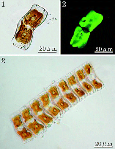
Figs 1–3. LMs of living cells showing chloroplasts. Fig. 1. Girdle view of single cell showing two large plastids, one on either side of the median transapical plane, each with a rounded pyrenoid. Fig. 2. CLSM of individual in Fig. 1 showing autofluorescence of the two plastids. Fig. 3. Colony of nine cells. Colonies are usually epiphytic on filamentous algae and attached by a stalk secreted by the lowermost cell.

Figs 4–7. LMs of cleaned vegetative frustules showing valve and girdle morphology. . RV (a) and ARV (b) of the same individuals. shows a small cell at the lower end of the size range. shows a large cell near the upper end of the size range. Figs 6–7. Girdle views showing the convex ARV, and the concave RV. Scale bar applies to all figures.
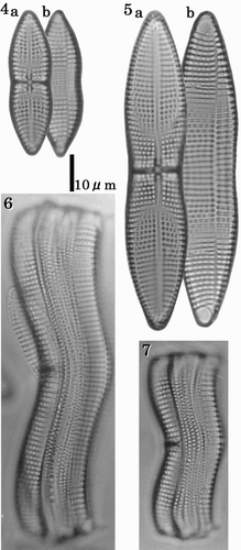
Figs 4–7. LMs of cleaned vegetative frustules showing valve and girdle morphology. Figs 4–5. RV (a) and ARV (b) of the same individuals. Fig. 4 shows a small cell at the lower end of the size range. Fig. 5 shows a large cell near the upper end of the size range. Figs 6–7. Girdle views showing the convex ARV, and the concave RV. Scale bar applies to all figures.

Figs 8–21. SEMs of vegetative cells. . External view of the RV showing filiform raphe fissure. . External view of RV centre showing the transverse stauros. . Terminal raphe fissure, curved slightly in the opposite direction to the central pores. . Internal view of the RV with valvocopula; costae are raised above the inner surface of the valve. . Internal view of cribra. . Internal view of centre of valve showing the thickened stauros. . Internal view of the terminal raphe fissure, which curves in the opposite direction to the external terminal raphe fissure. . External view of the ARV with marginal ridges and terminal spines. . Detail showing smooth edge of a marginal ridge. . Terminal spine and terminal orbiculus. Terminal spine emerges from the rapheless sternum. . Internal view of the ARV. . External view of cribra. . Internal view of cribra. Note that cribra are usually supported at 4 or 5 points. . Internal view of terminal orbiculus showing the occluding cribrum.
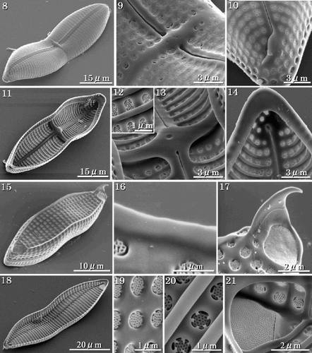
Figs 8–21. SEMs of vegetative cells. Fig. 8. External view of the RV showing filiform raphe fissure. Fig. 9. External view of RV centre showing the transverse stauros. Fig. 10. Terminal raphe fissure, curved slightly in the opposite direction to the central pores. Fig. 11. Internal view of the RV with valvocopula; costae are raised above the inner surface of the valve. Fig. 12. Internal view of cribra. Fig. 13. Internal view of centre of valve showing the thickened stauros. Fig. 14. Internal view of the terminal raphe fissure, which curves in the opposite direction to the external terminal raphe fissure. Fig. 15. External view of the ARV with marginal ridges and terminal spines. Fig. 16. Detail showing smooth edge of a marginal ridge. Fig. 17. Terminal spine and terminal orbiculus. Terminal spine emerges from the rapheless sternum. Fig. 18. Internal view of the ARV. Fig. 19. External view of cribra. Fig. 20. Internal view of cribra. Note that cribra are usually supported at 4 or 5 points. Fig. 21. Internal view of terminal orbiculus showing the occluding cribrum.

Figs 22–26. SEMs of vegetative frustules. . Enlarged view of the connection between adjacent individuals in apical terminal view. The terminal spine is used in aligning the colony. . Girdle view. . Valvocopula of the RV with two rows of areolae, one with roundish quadrangular areolae (arrowhead), the other with rectangular areolae (double-arrowhead). . Open ends of a girdle band. . Opposite closed end of the band shown in .
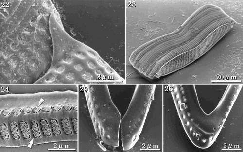
Figs 22–26. SEMs of vegetative frustules. Fig. 22. Enlarged view of the connection between adjacent individuals in apical terminal view. The terminal spine is used in aligning the colony. Fig. 23. Girdle view. Fig. 24. Valvocopula of the RV with two rows of areolae, one with roundish quadrangular areolae (arrowhead), the other with rectangular areolae (double-arrowhead). Fig. 25. Open ends of a girdle band. Fig. 26. Opposite closed end of the band shown in Fig. 25.

Figs 27–29. LMs of auxospores and initial cells. Arrowheads indicate mother frustules. . Auxospores attached to one of the mother frustules. . Perizonia of pre-initial cells that have not formed RVs, ARVs or girdles. . Initial cells. Note that these become about three times the size of the mother cell.
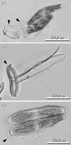
Figs 27–29. LMs of auxospores and initial cells. Arrowheads indicate mother frustules. Fig. 27. Auxospores attached to one of the mother frustules. Fig. 28. Perizonia of pre-initial cells that have not formed RVs, ARVs or girdles. Fig. 29. Initial cells. Note that these become about three times the size of the mother cell.

Figs 30–37. SEMs of pre-initial and initial cells. . Pre-initial cells. . Immediately after formation, initial valve enveloped by large amounts of mucilage. . RV of the initial cell showing the RV covered with a longitudinal perizonium. . Internal view of the longitudinal perizonium. . External view of ARV of an initial cell with a rounded valve face, orbiculi and terminal spines. . Portion of initial ARV showing rapheless sternum and cribra. . Terminal orbiculus. Orbiculi of initial cells are similar to those in the vegetative valves, however they often lack terminal spines. . Internal view of ARV of the initial cell. The rapheless sternum lies along the centre of the valve.
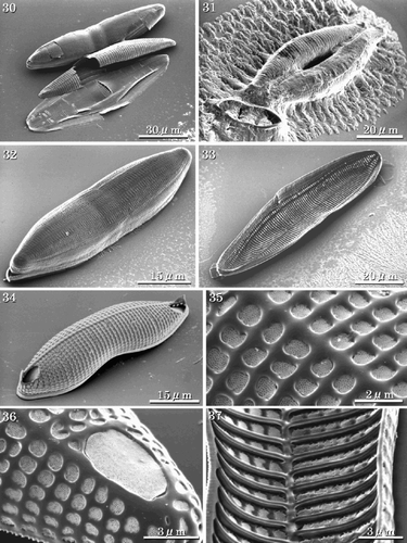
Figs 30–37. SEMs of pre-initial and initial cells. Fig. 30. Pre-initial cells. Fig. 31. Immediately after formation, initial valve enveloped by large amounts of mucilage. Fig. 32. RV of the initial cell showing the RV covered with a longitudinal perizonium. Fig. 33. Internal view of the longitudinal perizonium. Fig. 34. External view of ARV of an initial cell with a rounded valve face, orbiculi and terminal spines. Fig. 35. Portion of initial ARV showing rapheless sternum and cribra. Fig. 36. Terminal orbiculus. Orbiculi of initial cells are similar to those in the vegetative valves, however they often lack terminal spines. Fig. 37. Internal view of ARV of the initial cell. The rapheless sternum lies along the centre of the valve.

Figs 38–44. SEMs and TEMs showing perizonial structure. . SEM of early stage perizonia showing concave large perizonial bands. . Detail of , arrow indicating the growing edge of the perizonial band. . TEM of the fully formed perizonium composed of one large longitudinal band and four closed bands. . Detail of showing rows of areolae in the closed bands. . Detail of showing areolae occluded by cribra. . SEM of longitudinal perizonium. . Detail of the centre of the perizonium, with a break across the longitudinal band revealing a sternum-like thickening (double arrowhead). Abbreviations: P1: central longitudinal large band; P2: second closed band; P3: third closed band; P4: fourth closed band; P5: the final closed band.
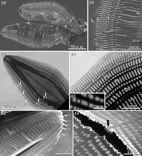
Figs 38–44. SEMs and TEMs showing perizonial structure. Fig. 38. SEM of early stage perizonia showing concave large perizonial bands. Fig. 39. Detail of Fig. 38, arrow indicating the growing edge of the perizonial band. Fig. 40. TEM of the fully formed perizonium composed of one large longitudinal band and four closed bands. Fig. 41. Detail of Fig. 40 showing rows of areolae in the closed bands. Fig. 42. Detail of Fig. 41 showing areolae occluded by cribra. Fig. 43. SEM of longitudinal perizonium. Fig. 44. Detail of the centre of the perizonium, with a break across the longitudinal band revealing a sternum-like thickening (double arrowhead). Abbreviations: P1: central longitudinal large band; P2: second closed band; P3: third closed band; P4: fourth closed band; P5: the final closed band.

Figs 45–47. Structure of initial cells (SEM and FIB). . Initial cells before sectioning. Grey rectangle denotes location of cross section. . Initial cells after sectioning. . Section of initial valves showing the RVs, ARVs, girdle bands and longitudinal perizonium. Abbreviations: P1: central longitudinal perizonial band; P2–P5: closed perizonial bands; RF: raphe fissure; VC: valvocopula; GB: girdle bands.
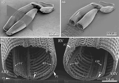
Figs 45–47. Structure of initial cells (SEM and FIB). Fig. 45. Initial cells before sectioning. Grey rectangle denotes location of cross section. Fig. 46. Initial cells after sectioning. Fig. 47. Section of initial valves showing the RVs, ARVs, girdle bands and longitudinal perizonium. Abbreviations: P1: central longitudinal perizonial band; P2–P5: closed perizonial bands; RF: raphe fissure; VC: valvocopula; GB: girdle bands.

Reprints and Corporate Permissions

Please note: Selecting permissions does not provide access to the full text of the article, please see our help page How do I view content?

To request a reprint or corporate permissions for this article, please click on the relevant link below:

Academic Permissions

Please note: Selecting permissions does not provide access to the full text of the article, please see our help page How do I view content?

Obtain permissions instantly via Rightslink by clicking on the button below:

If you are unable to obtain permissions via Rightslink, please complete and submit this Permissions form. For more information, please visit our Permissions help page.