797
Views
15
CrossRef citations to date
0
Altmetric
Original Articles

Vegetative and reproductive morphology of Gloiocladia repens (C. Agardh) Sánchez et Rodríguez-Prieto comb. nov. (Rhodymeniales, Rhodophyta), with a taxonomic re-assessment of the genera Fauchea and Gloiocladia

, &
Pages 145-162 | Received 02 Jul 2006, Accepted 08 Jan 2007, Published online: 18 May 2007

Figures & data

Figs 1–7. Gloiocladia repens. Habit. . Habit of the lectotype (LD 25705). . Habit of a fertile male and female gametophyte in the original collection (LD 25704). . Habit of a sterile specimen in the original collection (LD 25702). . Habit of a fertile female gametophyte in the original collection (LD 25703). . Habit of a tetrasporophyte (HGI-A 6320). . Habit of a sterile stipitate specimen (HGI-A 6746). . Detail of the thallus showing marginal haptera and thalli anastomoses (HGI-A 5463). Abbreviations: a: anastomosis; dh: discoid holdfast; h: hapterum; st: stipe. Scale bars: 1 cm (Figs 1–6); 0.5 cm (Fig. 7).
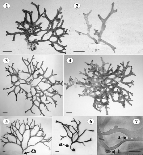
Figs 1–7. Gloiocladia repens. Habit. Fig. 1. Habit of the lectotype (LD 25705). Fig. 2. Habit of a fertile male and female gametophyte in the original collection (LD 25704). Fig. 3. Habit of a sterile specimen in the original collection (LD 25702). Fig. 4. Habit of a fertile female gametophyte in the original collection (LD 25703). Fig. 5. Habit of a tetrasporophyte (HGI-A 6320). Fig. 6. Habit of a sterile stipitate specimen (HGI-A 6746). Fig. 7. Detail of the thallus showing marginal haptera and thalli anastomoses (HGI-A 5463). Abbreviations: a: anastomosis; dh: discoid holdfast; h: hapterum; st: stipe. Scale bars: 1 cm (Figs 1–6); 0.5 cm (Fig. 7).

Table 1. Gloiocladia repens. Examined specimens. Locality, legit, date and depth of collection, herbarium identification number and phenology are indicated. All the specimens come from the coasts of Spain

Figs 8–13. Gloiocladia repens. Vegetative structure. Aniline blue staining. . Longitudinal (8) and transverse (9) sections of a sterile individual in the middle part of the plant (HGI-A 5631, 5637). . Cortex and subcortex in longitudinal (10) and transverse (11) section (HGI-A 5631, 5637). . Outer cortical cells in a surface view (HGI-A 5629). . Network of subcortical cells showing frequent secondary pit-connections between cells (HGI-A 5637). Abbreviation: spc: secondary pit-connection. Scale bars: 50 µm.
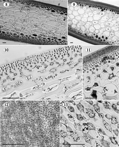
Figs 8–13. Gloiocladia repens. Vegetative structure. Aniline blue staining. Figs 8, 9. Longitudinal (8) and transverse (9) sections of a sterile individual in the middle part of the plant (HGI-A 5631, 5637). Figs 10, 11. Cortex and subcortex in longitudinal (10) and transverse (11) section (HGI-A 5631, 5637). Fig. 12. Outer cortical cells in a surface view (HGI-A 5629). Fig. 13. Network of subcortical cells showing frequent secondary pit-connections between cells (HGI-A 5637). Abbreviation: spc: secondary pit-connection. Scale bars: 50 µm.

Figs 14–18. Gloiocladia repens. Vegetative structure. Aniline blue staining. . Medullary cells in transverse section showing several secondary pit-connections between them (HGI-A 5463). . Transverse section of the basal part of the thallus showing the development of rhizoidal filaments and secondary medullary cells (HGI-A 5632). . Detail of the rhizoidal filaments connected by secondary pit-connections (HGI-A 5632). . Transverse section of the basal part of the thallus with many secondary medullary cells (HGI-A 5632). . Transverse section of the stipe (HGI-A 1793). Abbreviations: rf: rhizoidal filament; smc: secondary medullary cell; spc: secondary pit-connection. Scale bars: 50 µm (Fig. 14); 100 µm (Figs 15, 17, 18); 25 µm (Fig. 16).
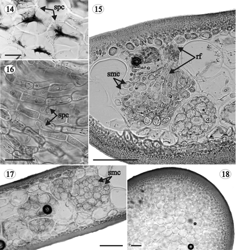
Figs 14–18. Gloiocladia repens. Vegetative structure. Aniline blue staining. Fig. 14. Medullary cells in transverse section showing several secondary pit-connections between them (HGI-A 5463). Fig. 15. Transverse section of the basal part of the thallus showing the development of rhizoidal filaments and secondary medullary cells (HGI-A 5632). Fig. 16. Detail of the rhizoidal filaments connected by secondary pit-connections (HGI-A 5632). Fig. 17. Transverse section of the basal part of the thallus with many secondary medullary cells (HGI-A 5632). Fig. 18. Transverse section of the stipe (HGI-A 1793). Abbreviations: rf: rhizoidal filament; smc: secondary medullary cell; spc: secondary pit-connection. Scale bars: 50 µm (Fig. 14); 100 µm (Figs 15, 17, 18); 25 µm (Fig. 16).

Figs 19, 20. Gloiocladia repens. Light photomicrographs of male reproductive structures. Aniline blue staining. . Transverse section of a fertile area of the thallus showing a spermatangial sorus (HGI-A 6303). . Transverse section of a fertile area of the thallus showing spermatia borne on spermatangial mother cells (LD 25704). Abbreviations: cc: cortical cell, s: spermatium; spmc: spermatangial mother cell. Scale bars: 50 µm.
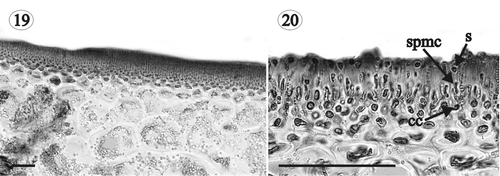
Figs 19, 20. Gloiocladia repens. Light photomicrographs of male reproductive structures. Aniline blue staining. Fig. 19. Transverse section of a fertile area of the thallus showing a spermatangial sorus (HGI-A 6303). Fig. 20. Transverse section of a fertile area of the thallus showing spermatia borne on spermatangial mother cells (LD 25704). Abbreviations: cc: cortical cell, s: spermatium; spmc: spermatangial mother cell. Scale bars: 50 µm.

Figs 21–25. Gloiocladia repens. Light photomicrographs of female reproductive structures and postfertilization stages. Aniline blue staining. . Carpogonial branch (HGI-A 6303). . Procarp at a stage in which the first cell of the auxiliary branch can just be distinguished (HGI-A 6303). . Auxiliary cell branch composed of an auxiliary mother cell and an auxiliary cell with a proteinaceous inclusion (HGI-A 6334). . Young gonimoblast arising from the primary gonimoblast cell. The fusion cell between the carpogonial branch cells can also been seen (LD 25703). . Detail of the fusion cell (HGI-A 6303). Abbreviations: ac: auxiliary cell; amc: auxiliary mother cell; c: carposporangia; cbc: cells of the carpogonial branch; cc: cortical cell; cp: carpogonium; fc: fusion cell; fcab: first cell of the auxiliary branch; fcbc: fused cells of the carpogonial branch; g: gonimoblast; nc: nutritive cell; pgc: primary gonimoblast cell; pi: proteinaceous inclusion; sc: supporting cell; tr: trichogyne; stc: sterile cell; tac: cell of the tela arachnoidea. Scale bars: 25 µm (Figs 21, 22); 50 µm (Figs 23–25).
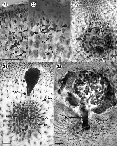
Figs 21–25. Gloiocladia repens. Light photomicrographs of female reproductive structures and postfertilization stages. Aniline blue staining. Fig. 21. Carpogonial branch (HGI-A 6303). Fig. 22. Procarp at a stage in which the first cell of the auxiliary branch can just be distinguished (HGI-A 6303). Fig. 23. Auxiliary cell branch composed of an auxiliary mother cell and an auxiliary cell with a proteinaceous inclusion (HGI-A 6334). Fig. 24. Young gonimoblast arising from the primary gonimoblast cell. The fusion cell between the carpogonial branch cells can also been seen (LD 25703). Fig. 25. Detail of the fusion cell (HGI-A 6303). Abbreviations: ac: auxiliary cell; amc: auxiliary mother cell; c: carposporangia; cbc: cells of the carpogonial branch; cc: cortical cell; cp: carpogonium; fc: fusion cell; fcab: first cell of the auxiliary branch; fcbc: fused cells of the carpogonial branch; g: gonimoblast; nc: nutritive cell; pgc: primary gonimoblast cell; pi: proteinaceous inclusion; sc: supporting cell; tr: trichogyne; stc: sterile cell; tac: cell of the tela arachnoidea. Scale bars: 25 µm (Figs 21, 22); 50 µm (Figs 23–25).

Figs 26–30. Gloiocladia repens. Light photomicrographs of the female reproductive structures with aniline blue staining. . Detail of stellate nutritive cells (HGI-A 5542). . Details of stellate cells of the tela arachnoidea (HGI-A 5542). . Cystocarps (LD 25703). . Detail of the carposporangia going out through the ostiole (HGI-A 5542). Scale bars: 50 µm (Figs 26–28, 30); 5 mm (Fig. 29).
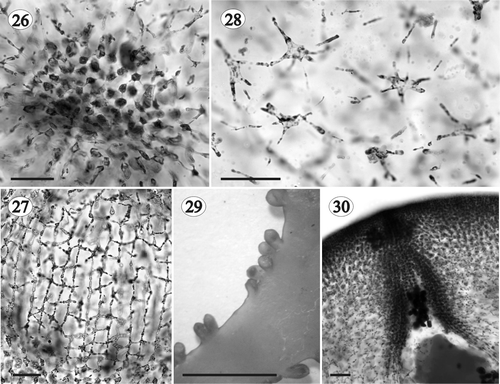
Figs 26–30. Gloiocladia repens. Light photomicrographs of the female reproductive structures with aniline blue staining. Fig. 26. Detail of stellate nutritive cells (HGI-A 5542). Figs 27, 28. Details of stellate cells of the tela arachnoidea (HGI-A 5542). Fig. 29. Cystocarps (LD 25703). Fig. 30. Detail of the carposporangia going out through the ostiole (HGI-A 5542). Scale bars: 50 µm (Figs 26–28, 30); 5 mm (Fig. 29).

Figs 31–35. Gloiocladia repens. Tetrasporangial development. . Nemathecia in branch surface (HGI-A 6320). . Transverse section of a well-developed nemathecium (HGI-A 5463). . Transverse section of nemathecium with cruciately dividing tetrasporangia (HGI-A 6298). . Transverse section of nemathecium with decussately cruciate dividing tetrasporangia (HGI-A 6302) (aniline blue staining). . Transverse section of nemathecium with irregularly dividing tetrasporangia (HGI-A 6302) (aniline blue staining). Scale bars: 5 mm (Fig. 31); 100 µm (Fig. 32); 25 µm (Figs 33–35).
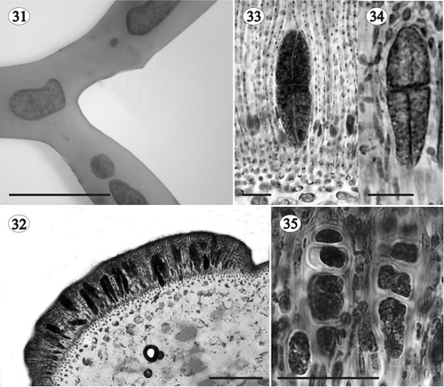
Figs 31–35. Gloiocladia repens. Tetrasporangial development. Fig. 31. Nemathecia in branch surface (HGI-A 6320). Fig. 32. Transverse section of a well-developed nemathecium (HGI-A 5463). Fig. 33. Transverse section of nemathecium with cruciately dividing tetrasporangia (HGI-A 6298). Fig. 34. Transverse section of nemathecium with decussately cruciate dividing tetrasporangia (HGI-A 6302) (aniline blue staining). Fig. 35. Transverse section of nemathecium with irregularly dividing tetrasporangia (HGI-A 6302) (aniline blue staining). Scale bars: 5 mm (Fig. 31); 100 µm (Fig. 32); 25 µm (Figs 33–35).

Figs 36, 37. Gloiocladia repens and Gloiocladia furcata. Tetrasporangial pit-connections with aniline blue staining. . Tetrasporangia of G. repens showing basal pit-connection with cortical filaments (HGI-A 5631). . Tetrasporangia of G. furcata showing lateral pit-connection with cortical filaments (HGI-A 5769). Abbreviations: bpc: basal pit-connection; lpc: lateral pit-connection; t: tetrasporangium; yt: young tetrasporangium. Scale bars: 50 µm.
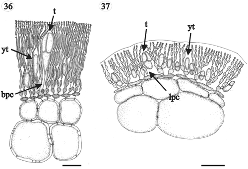
Figs 36, 37. Gloiocladia repens and Gloiocladia furcata. Tetrasporangial pit-connections with aniline blue staining. Fig. 36. Tetrasporangia of G. repens showing basal pit-connection with cortical filaments (HGI-A 5631). Fig. 37. Tetrasporangia of G. furcata showing lateral pit-connection with cortical filaments (HGI-A 5769). Abbreviations: bpc: basal pit-connection; lpc: lateral pit-connection; t: tetrasporangium; yt: young tetrasporangium. Scale bars: 50 µm.

Fig. 38. Phylogenetic trees resulting from analyses of 18S rRNA gene sequences of Faucheacean species. Accession numbers for previously published sequences are given below species names. Sequences generated in this study are shown in larger font. (A) One of four most parsimonious trees (length = 176; CI = 0.687; RI = 0.580). Inferred branch lengths are shown above branches, parsimony (P) and distance (D) bootstrap proportions are shown below branches. (B) Maximum-likelihood tree (lnL = −3340.58416) with likelihood bootstrap proportions shown above branches.

Fig. 38. Phylogenetic trees resulting from analyses of 18S rRNA gene sequences of Faucheacean species. Accession numbers for previously published sequences are given below species names. Sequences generated in this study are shown in larger font. (A) One of four most parsimonious trees (length = 176; CI = 0.687; RI = 0.580). Inferred branch lengths are shown above branches, parsimony (P) and distance (D) bootstrap proportions are shown below branches. (B) Maximum-likelihood tree (lnL = −3340.58416) with likelihood bootstrap proportions shown above branches.

Table 2. Comparison of the main features of species currently placed in the genus Fauchea and transferred here to Gloiocladia

Table 3. Formal transfers to the genus Gloiocladia

Reprints and Corporate Permissions

Please note: Selecting permissions does not provide access to the full text of the article, please see our help page How do I view content?

To request a reprint or corporate permissions for this article, please click on the relevant link below:

Academic Permissions

Please note: Selecting permissions does not provide access to the full text of the article, please see our help page How do I view content?

Obtain permissions instantly via Rightslink by clicking on the button below:

If you are unable to obtain permissions via Rightslink, please complete and submit this Permissions form. For more information, please visit our Permissions help page.