Figures & data
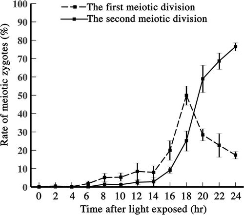
Fig. 1. Proportions of zygotes completing the first and second meiotic divisions, counted after exposing the zygotes to light. Dashed line: zygotes completing the first meiotic division. Solid line: zygotes completing the second meiotic division. Vertical line: standard deviation.
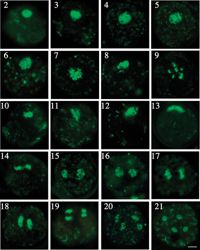
Figs 2–21. Fluorescence microscopy of chromosome behaviour during meiosis. . Chromosome morphology in prophase I. . Leptotene. . Zygotene. . Pachytene. . Diplotene and diakinesis at approximately 16 h. . Metaphase I to telophase II. . Metaphase I. . Anaphase I. . Telophase I. . The side view shows chromosomes situated at the surface of the zygotes. . Prophase II. . Metaphase II. . Anaphase II. . Telophase II. . Formation of four daughter cells. Scale bar: 5 µm.
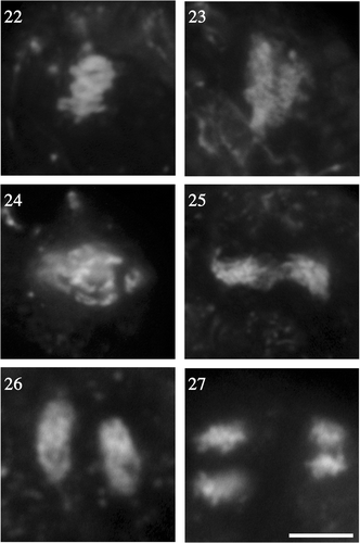
Figs 22–27. Enlargements of chromosomes at metaphase and anaphase. . Chromosomes at meiosis I, as shown in fluorescent photomicrographs of enlarged portions of the nuclei in , respectively. . Chromosomes at meiosis II (enlarged portions of the nuclei in ). Scale bar: 5 µm.
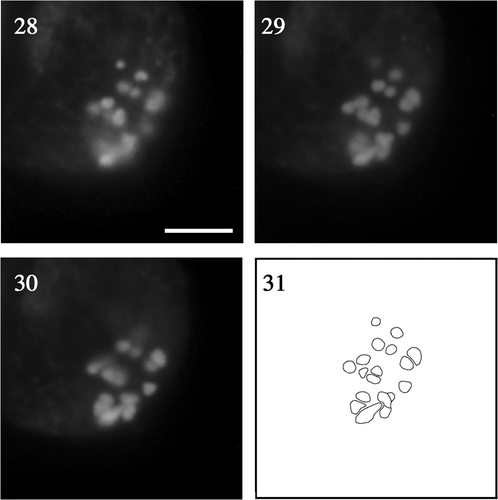
Figs 28–31. Fluorescence micrographs of chromosomes at meiotic diakinesis at different focal planes of the same zygote. . Low. . Middle. . Upper. . Composite image based on the superimposition of views . Scale bar: 5 µm.
Figs 32–35. Fluorescent photomicrographs of spread chromosomes at diakinesis. . Total chromosomes spread on glass slide. . Chromosomes arranged in order of fluorescence intensity. Scale bar: 5 µm.
