1,223
Views
8
CrossRef citations to date
0
Altmetric
Original Articles

Observations on the diatom Navicula hedinii Hustedt (Bacillariophyceae) and its transfer to a new genus Envekadea Van de Vijver et al. gen. nov.

, , , , , , & show all
Pages 123-138 | Received 29 Oct 2007, Accepted 16 Jun 2008, Published online: 12 Mar 2009

Figures & data

Figs 1–16. Light micrographs of Envekadea (Navicula) hedinii. . Valve views of specimens from Lake Vrana (Croatia). . Girdle view of specimen from Lake Vrana (Cr.). . Valve views of the type population from Mapiek Köll (eastern Turkestan). . Valve view of specimen (Croatian population) showing the chloroplast structure. Scale bars: 10 µm.
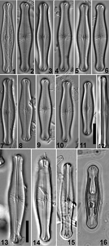
Figs 1–16. Light micrographs of Envekadea (Navicula) hedinii. Figs 1–11. Valve views of specimens from Lake Vrana (Croatia). Fig. 12. Girdle view of specimen from Lake Vrana (Cr.). Figs 13–15. Valve views of the type population from Mapiek Köll (eastern Turkestan). Fig. 16. Valve view of specimen (Croatian population) showing the chloroplast structure. Scale bars: 10 µm.

Figs 17–19. Light micrographs of valve views of Envekadea (Navicula) pseudocrassirostris from Eleusis (Athens, Greece). Scale bar: 10 µm.
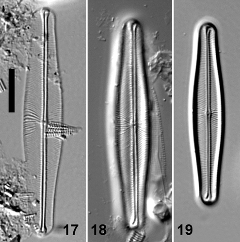
Figs 17–19. Light micrographs of valve views of Envekadea (Navicula) pseudocrassirostris from Eleusis (Athens, Greece). Scale bar: 10 µm.

Figs 20–22. Transmission electron micrographs of Envekadea (Navicula) hedinii from Lake Vrana (Croatia). . Entire valve showing the square to polygonal shape of the areolae when the external hymenes have been removed. . Detail of the stria structure with the hymenes covering the areolae. . Detail of the hymenes showing their porous structure. Scale bars: 10 µm (), 1 µm () and 0.5 µm ().
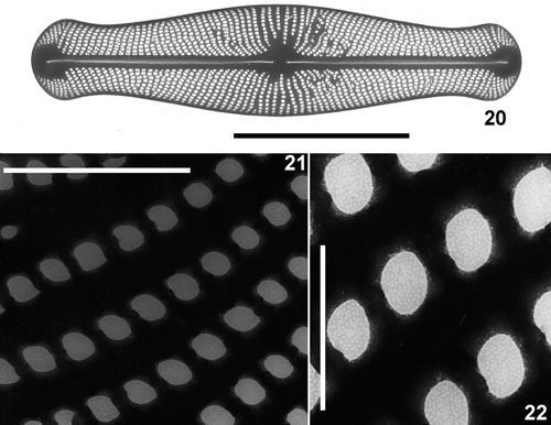
Figs 20–22. Transmission electron micrographs of Envekadea (Navicula) hedinii from Lake Vrana (Croatia). Fig. 20. Entire valve showing the square to polygonal shape of the areolae when the external hymenes have been removed. Fig. 21. Detail of the stria structure with the hymenes covering the areolae. Fig. 22. Detail of the hymenes showing their porous structure. Scale bars: 10 µm (Fig. 20), 1 µm (Fig. 21) and 0.5 µm (Fig. 22).

Figs 23–27. Scanning electron micrographs of external views of Envekadea (Navicula) hedinii from Lake Vrana (Croatia). . Presence of the hymenes covering the external areola openings. . An eroded valve showing the raphe path with the fissures deflected into opposite directions and the typical areola structure. . Detail of the terminal raphe fissure. Note the almost straight course of the raphe branch and the irregular areola structure. . Detail of the delta-shaped central raphe pores. . A tilted valve showing the stria structure at the valve face/mantle junction. Scale bars: 10 µm () and 1 µm ().
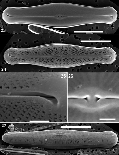
Figs 23–27. Scanning electron micrographs of external views of Envekadea (Navicula) hedinii from Lake Vrana (Croatia). Fig. 23. Presence of the hymenes covering the external areola openings. Fig. 24. An eroded valve showing the raphe path with the fissures deflected into opposite directions and the typical areola structure. Fig. 25. Detail of the terminal raphe fissure. Note the almost straight course of the raphe branch and the irregular areola structure. Fig. 26. Detail of the delta-shaped central raphe pores. Fig. 27. A tilted valve showing the stria structure at the valve face/mantle junction. Scale bars: 10 µm (Figs 23, 24, 27) and 1 µm (Figs 25, 26).

Figs 28–33. Scanning electron micrographs of valve and girdle views of Envekadea (Navicula) hedinii from Lake Vrana (Croatia). . Detail of the external valve apex with the wedge-shaped hyaline area and the clearly deflected terminal raphe fissure. . External detail of the irregularly shaped areolae near the valve apex. . Internal view of an entire valve with a partly detached valvocopula lying in the valve. . Detail of the external valve apex showing the girdle. . Detail of the valvocopula in , showing the lack of perforations and the undulated pars interior. . End of a frustule showing girdle bands and valve apices. Scale bars: 10 µm () and 1 µm ().
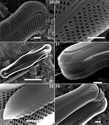
Figs 28–33. Scanning electron micrographs of valve and girdle views of Envekadea (Navicula) hedinii from Lake Vrana (Croatia). Fig. 28. Detail of the external valve apex with the wedge-shaped hyaline area and the clearly deflected terminal raphe fissure. Fig. 29. External detail of the irregularly shaped areolae near the valve apex. Fig. 30. Internal view of an entire valve with a partly detached valvocopula lying in the valve. Fig. 31. Detail of the external valve apex showing the girdle. Fig. 32. Detail of the valvocopula in Fig. 30, showing the lack of perforations and the undulated pars interior. Fig. 33. End of a frustule showing girdle bands and valve apices. Scale bars: 10 µm (Fig. 30) and 1 µm (Figs 28, 29, 31–33).

Figs 34–38. Scanning electron micrographs of internal views of Envekadea (Navicula) hedinii from Lake Vrana (Croatia). . Entire valve. . Detail of the central part of the valve with the raised internal nodule. Note the raised rims between the striae and the round shape of the areolae. . Detail of the central raphe endings. Note the slightly deflected ends. . Detail of the valve apex with the typical, very simple helictoglossa. . Detail of the valve apex with the typical, tunnel-like helictoglossa. Scale bars: 10 µm () and 1 µm ().
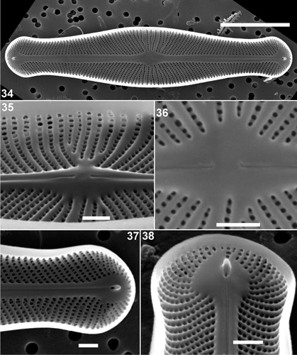
Figs 34–38. Scanning electron micrographs of internal views of Envekadea (Navicula) hedinii from Lake Vrana (Croatia). Fig. 34. Entire valve. Fig. 35. Detail of the central part of the valve with the raised internal nodule. Note the raised rims between the striae and the round shape of the areolae. Fig. 36. Detail of the central raphe endings. Note the slightly deflected ends. Fig. 37. Detail of the valve apex with the typical, very simple helictoglossa. Fig. 38. Detail of the valve apex with the typical, tunnel-like helictoglossa. Scale bars: 10 µm (Fig. 34) and 1 µm (Figs 35–38).

Figs 39–44. Scanning electron micrographs of Envekadea (Navicula) hedinii from the type population of Mapiek Köll. . External valve view of an entire valve. Presence of the hymenes covering the areola openings. . Detail of the central part of the valve showing the central raphe endings. . Detail of an external valve apex with the typical, deflected terminal raphe fissure. . Internal view of an entire valve. Although broken along the axial area, it is still clear that the central raphe endings are weakly deflected. . Detail of the internal structure of a valve apex with the simple helictoglossa. The presence of the raised rims between the striae and the rounded shape of the areolae is well visible. . Detail of a broken valve showing the presence of external hymenes covering the external areola openings. Inside the valve, it is clear that internal hymenes are completely absent. Scale bars: 10 µm () and 5 µm ().
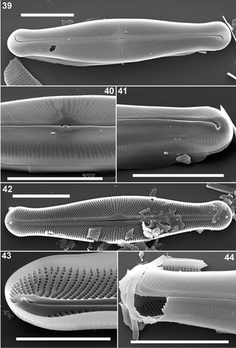
Figs 39–44. Scanning electron micrographs of Envekadea (Navicula) hedinii from the type population of Mapiek Köll. Fig. 39. External valve view of an entire valve. Presence of the hymenes covering the areola openings. Fig. 40. Detail of the central part of the valve showing the central raphe endings. Fig. 41. Detail of an external valve apex with the typical, deflected terminal raphe fissure. Fig. 42. Internal view of an entire valve. Although broken along the axial area, it is still clear that the central raphe endings are weakly deflected. Fig. 43. Detail of the internal structure of a valve apex with the simple helictoglossa. The presence of the raised rims between the striae and the rounded shape of the areolae is well visible. Fig. 44. Detail of a broken valve showing the presence of external hymenes covering the external areola openings. Inside the valve, it is clear that internal hymenes are completely absent. Scale bars: 10 µm (Figs 39–42, 44) and 5 µm (Fig. 43).

Figs. 45–50. Scanning electron micrographs of Envekadea (Navicula) pseudocrassirostris from Eleusis (Athens, Greece). . External view of an entire valve in a slightly tilted position to show the valve face/mantle margin. Note the sigmoid course of the raphe. . Detail of the external central area with the delta-shaped central raphe pores and the reduced central area. . Detail of an external valve apex with the golfclub-like terminal fissures. The internal raphe branch does not seem to follow the terminal fissure path. . Internal view of an entire valve. . Detail of the girdle. Three unperforated bands can be seen. . Detail of the internal structure of the apex with the simple helictoglossa and the wedge-shaped hyaline area around the terminal ending. Scale bars represent 10 µm () and 1 µm ().
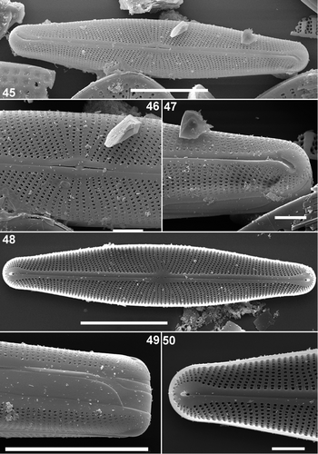
Figs. 45–50. Scanning electron micrographs of Envekadea (Navicula) pseudocrassirostris from Eleusis (Athens, Greece). Fig. 45. External view of an entire valve in a slightly tilted position to show the valve face/mantle margin. Note the sigmoid course of the raphe. Fig. 46. Detail of the external central area with the delta-shaped central raphe pores and the reduced central area. Fig. 47. Detail of an external valve apex with the golfclub-like terminal fissures. The internal raphe branch does not seem to follow the terminal fissure path. Fig. 48. Internal view of an entire valve. Fig. 49. Detail of the girdle. Three unperforated bands can be seen. Fig. 50. Detail of the internal structure of the apex with the simple helictoglossa and the wedge-shaped hyaline area around the terminal ending. Scale bars represent 10 µm (Figs 45, 48, 49) and 1 µm (Figs 46, 47, 50).

Table 1. Comparison between the new genus Envekadea and some freshwater genera presenting a similar set of features.

Table 2. Comparison between the new genus Envekadea and some marine genera presenting a similar set of features.

Reprints and Corporate Permissions

Please note: Selecting permissions does not provide access to the full text of the article, please see our help page How do I view content?

To request a reprint or corporate permissions for this article, please click on the relevant link below:

Academic Permissions

Please note: Selecting permissions does not provide access to the full text of the article, please see our help page How do I view content?

Obtain permissions instantly via Rightslink by clicking on the button below:

If you are unable to obtain permissions via Rightslink, please complete and submit this Permissions form. For more information, please visit our Permissions help page.