Figures & data
Figs 1–9. SEM micrographs of the cyst of Peridinium umbonatum sensu lato. . Cyst with no thecal remains seen in apical view in which the slightly flattened ventral side is visible and the paratabulation by parasutures is discernable. . Cyst partially covered in thecal vestiges seen in dorsal view. . Cyst partially covered in thecal vestiges seen in ventral view. . Detail of showing the apical pore (ap) region with the x-plate (*). . Cysts with no thecal remains in which the paratabulation is discernable by parasutures. . The ventral-dorsal side of the cyst with the first antapical, polygonal, lightly peanut-shaped archeopyle. Key: X′: apical plates; Xa: intercalary plates; X″: precingular plates. Scale bars: 10 µm.

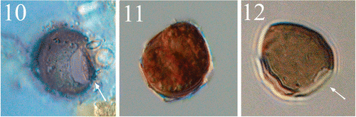
Figs 10–12. Light micrographs of the cyst of Peridinium umbonatum sensu lato. . Antapical view of an empty cyst with the operculum (arrow) attached at the ventral side. . Full cyst. . Empty cyst with the archeopyle (arrow) detectable in the antapical position.
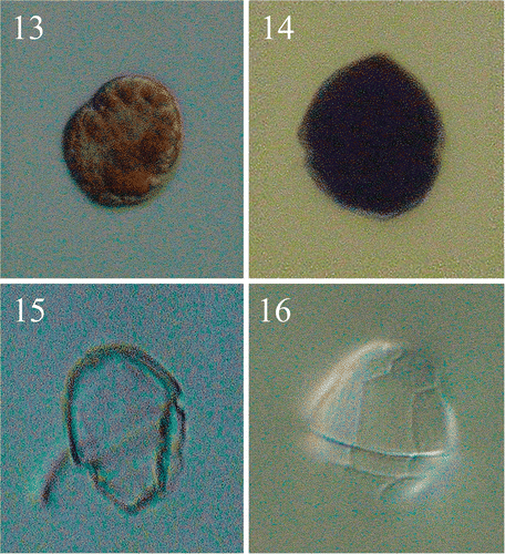
Figs 13–16. Light micrographs of the vegetative cells of Peridinium umbonatum sensu lato obtained from germinated cysts. . Non-thecated stage cell in which cingular portion is visible and radially arranged chloroplasts detectable. . Thecated stage cell. . Empty theca in lateral view, showing the slanting down profile of the hypocone. . Empty theca in dorsal view, showing the longer 4″ plate and the ‘remotum’ tabulation pattern.
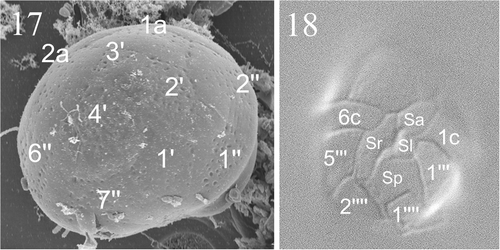
Figs 17, 18. Micrographs showing tabulation of cyst and vegetative cell. . SEM of a cyst of Peridinium umbonatum sensu lato showing details of the epicyst paratabulation. . LM showing the tabulation of the ventral side of the vegetative cell. Key: X′: apical plates; Xa: intercalary plates; X″: precingular plates; Xc: cingular plates; Sa: apical sulcal plate; Sp: posterior sulcal plate; Sr: right sulcal plate; Sl: left sulcal plate; X′″: postcingular plates; X″″: antapical plates).
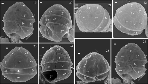
Figs 19–26. SEMs of the vegetative cells of Peridinium umbonatum sensu lato collected in the water column of Lake Nero di Cornisello. Key: X′: apical plates; Xa: intercalary plates; X″: precingular plates; Xc: cingular plates; X′″: postcingular plates. Scale bars: 1 µm.
Figs 27–29. Drawings of the plate pattern of Peridinium umbonatum sensu lato of Lake Nero di Cornisello in the apical (), dorsal (), and ventral () sides. Key: ap: apical pore; *: x-plate; X′: apical plates; Xa: intercalary plates; X″: precingular plates; Xc: cingular plates; Sa: apical sulcal plate; Sp: posterior sulcal plate; Sr: right sulcal plate; Sl: left sulcal plate; X′″: postcingular plates; X″″: antapical plates.
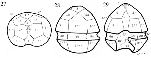
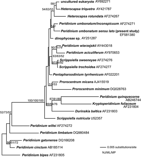
Fig. 30. Neighbor joining tree based on parts of the SSU the nuclear encoded rDNA. The tree is unrooted. The bootstrap values indicated on the branches represent bootstrap values from neighbor joining (500 replicates, before slash), (maximum likelihood 1000 replicates, between slashes) and parsimony (1000 replicates, after slash). Only values above 50% are shown.
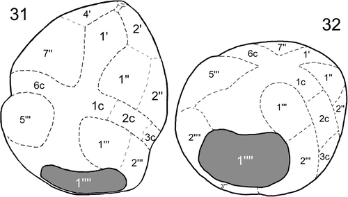
Figs 31, 32. Drawings of the cyst of Peridinium umbonatum sensu lato of Lake Nero di Cornisello illustrating the first antapical plate position (1″″) of the archeopyle in relation with the plate pattern of the thecate stage in the semi-ventral () and semi-antapical () side. Key: dashed lines: visible lines of paratabulation; dotted lines: interpreted lines of paratabulation; X′: apical plates; X″: precingular plates; Xc: cingular plates; X′″: postcingular plates; X″″: antapical plates).